1 list the main provisions of the cell theory. Cell as a biological system (multiple choice). What have we learned
Almost 400 years have passed since the discovery of cells, before the current state of the cell theory was formulated. For the first time a cell was investigated in 1665 by a naturalist from England. Having noticed cellular structures on a thin section of cork, he gave them the name of cells.
In his primitive microscope, Hooke could not yet see all the features, but as optical instruments improved, and methods for staining preparations appeared, scientists became more and more immersed in the world of fine cytological structures.
How did the cell theory come about?
A landmark discovery that influenced the further course of research and the current state of cell theory was made in the 30s of the 19th century. The Scot R. Brown, studying the leaf of a plant with a light microscope, found similar rounded seals in plant cells, which he later called nuclei.
From that moment, an important feature appeared for comparing the structural units of various organisms with each other, which became the basis for conclusions about the unity of the origin of the living. It is not for nothing that even the current position of cell theory contains a reference to this conclusion.

The question of the origin of cells was raised in 1838 by the German botanist Matthias Schleiden. Massively studying plant material, he noted that in all living plant tissues, the presence of nuclei is mandatory.
His compatriot zoologist Theodor Schwann made the same conclusions about animal tissue. Having studied the works of Schleiden and comparing many plant and animal cells, he concluded: despite the diversity, they all have a common feature - a formed nucleus.
The cell theory of Schwann and Schleiden
Having put together the available facts about the cell, T. Schwann and M. Schleiden put forward the main postulate. It was that all organisms (plants and animals) consist of cells that are similar in structure.

In 1858, another addition to the cell theory was made. proved that the body grows by increasing the number of cells by dividing the original maternal. It seems obvious to us, but for those times his discovery was very advanced and modern.
At that time, the current position of Schwann's cell theory in textbooks is formulated as follows:
- All tissues of living organisms have a cellular structure.
- Animal and plant cells are formed in the same way (cell division) and have a similar structure.
- The body consists of groups of cells, each of them is capable of independent life.
Having become one of the most important discoveries of the 19th century, the cell theory laid the foundation for the idea of the unity of origin and commonality of the evolutionary development of living organisms.
Further development of cytological knowledge
The improvement of research methods and equipment has allowed scientists to significantly deepen their knowledge of the structure and life of cells:
- the relationship between the structure and function of both individual organelles and cells as a whole (specialization of cytostructures) has been proven;
- each cell individually demonstrates all the properties inherent in living organisms (grows, reproduces, exchanges matter and energy with the environment, is mobile to one degree or another, adapts to changes, etc.);
- organelles cannot individually exhibit similar properties;
- in animals, fungi, plants, organelles identical in structure and function are found;
- All cells in the body are interconnected and work together to perform complex tasks.
Thanks to new discoveries, the provisions of the theory of Schwann and Schleiden were refined and supplemented. The modern scientific world uses the extended postulates of the fundamental theory in biology.
In the literature, you can find a different number of postulates of modern cell theory, the most complete version contains five points:
- The cell is the smallest (elementary) living system, the basis of the structure, reproduction, development and life of organisms. Non-cellular structures cannot be called living.
- Cells appear exclusively by dividing existing ones.
- The chemical composition and structure of the structural units of all living organisms are similar.
- A multicellular organism develops and grows by dividing one/several original cells.
- The similar cellular structure of the organisms inhabiting the Earth indicates a single source of their origin.

The original and modern provisions of the cell theory have much in common. Deep and extended postulates reflect the current level of knowledge on the structure, life and interaction of cells.
Theory for task 4 from the exam in biology
Cell as a biological system
Modern cellular theory, its main provisions, the role in the formation of the modern natural-science picture of the world. Development of knowledge about the cell. The cellular structure of organisms is the basis of the unity of the organic world, proof of the relationship of living nature
Modern cellular theory, its main provisions, role in the formation of the modern natural-science picture of the world
One of the fundamental concepts in modern biology is the idea that all living organisms have a cellular structure. Science deals with the study of the structure of the cell, its vital activity and interaction with the environment. cytology now commonly referred to as cell biology. Cytology owes its appearance to the formulation of the cellular theory (1838-1839, M. Schleiden, T. Schwann, supplemented in 1855 by R. Virchow).
cell theory is a generalized idea of the structure and functions of cells as living units, their reproduction and role in the formation of multicellular organisms.
The main provisions of the cell theory:
- A cell is a unit of structure, life activity, growth and development of living organisms - there is no life outside the cell.
- A cell is a single system consisting of many elements that are naturally connected with each other, representing a certain integral formation.
- The cells of all organisms are similar in their chemical composition, structure and functions.
- New cells are formed only as a result of division of mother cells (“cell from cell”).
- The cells of multicellular organisms form tissues, and organs are made up of tissues. The life of an organism as a whole is determined by the interaction of its constituent cells.
- The cells of multicellular organisms have a complete set of genes, but differ from each other in that different groups of genes work for them, which results in the morphological and functional diversity of cells - differentiation.
Thanks to the creation of the cellular theory, it became clear that the cell is the smallest unit of life, an elementary living system, which has all the signs and properties of living things. The formulation of the cell theory became the most important prerequisite for the development of views on heredity and variability, since the identification of their nature and their inherent patterns inevitably suggested the universality of the structure of living organisms. Revealing the unity of the chemical composition and structural plan of cells served as an impetus for the development of ideas about the origin of living organisms and their evolution. In addition, the origin of multicellular organisms from a single cell during embryonic development has become a dogma of modern embryology.
Development of knowledge about the cell
Until the 17th century, man knew nothing at all about the microstructure of the objects surrounding him and perceived the world with the naked eye. The instrument for studying the microcosm, the microscope, was invented approximately in 1590 by the Dutch mechanics G. and Z. Jansen, but its imperfection made it impossible to examine sufficiently small objects. Only the creation on its basis of the so-called compound microscope by K. Drebbel (1572-1634) contributed to the progress in this area.
In 1665, the English physicist R. Hooke (1635-1703) improved the design of the microscope and the technology of grinding lenses, and, wanting to make sure that the image quality improved, he examined sections of cork, charcoal and living plants under it. On the sections, he found the smallest pores resembling a honeycomb, and called them cells (from lat. cellula cell, cell). It is interesting to note that R. Hooke considered the cell membrane to be the main component of the cell.
In the second half of the 17th century, the works of the most prominent microscopists M. Malpighi (1628-1694) and N. Gru (1641-1712) appeared, who also discovered the cellular structure of many plants.
To make sure that what R. Hooke and other scientists saw was true, the Dutch merchant A. van Leeuwenhoek, who did not have a special education, independently developed a microscope design that was fundamentally different from the existing one, and improved the lens manufacturing technology. This allowed him to achieve an increase of 275-300 times and to consider such details of the structure that were technically inaccessible to other scientists. A. van Leeuwenhoek was an unsurpassed observer: he carefully sketched and described what he saw under a microscope, but did not seek to explain it. He discovered unicellular organisms, including bacteria, found nuclei, chloroplasts, thickenings of cell walls in plant cells, but his discoveries could be evaluated much later.
Discoveries of the components of the internal structure of organisms in the first half of the 19th century followed one after another. G. Mol distinguished in plant cells living matter and a watery liquid - cell sap, discovered pores. The English botanist R. Brown (1773-1858) discovered the nucleus in orchid cells in 1831, then it was found in all plant cells. The Czech scientist J. Purkinje (1787-1869) introduced the term "protoplasm" (1840) to refer to the semi-liquid gelatinous contents of a cell without a nucleus. The Belgian botanist M. Schleiden (1804-1881) advanced further than all his contemporaries, who, studying the development and differentiation of various cellular structures of higher plants, proved that all plant organisms originate from one cell. He also considered rounded nucleolus bodies in the nuclei of onion scale cells (1842).
In 1827, the Russian embryologist K. Baer discovered the eggs of humans and other mammals, thereby refuting the notion of the development of an organism exclusively from male gametes. In addition, he proved the formation of a multicellular animal organism from a single cell - a fertilized egg, as well as the similarity of the stages of embryonic development of multicellular animals, which suggested the unity of their origin. The information accumulated by the middle of the 19th century required generalization, which became the cellular theory. Biology owes its formulation to the German zoologist T. Schwann (1810-1882), who, based on his own data and M. Schleiden's conclusions on the development of plants, suggested that if a nucleus is present in any formation visible under a microscope, then this formation is cell. Based on this criterion, T. Schwann formulated the main provisions of the cell theory.
The German physician and pathologist R. Virchow (1821-1902) introduced another important provision into this theory: cells arise only by dividing the original cell, that is, cells are formed only from cells (“cell from cell”).
Since the creation of the cell theory, the doctrine of the cell as a unit of the structure, function and development of the organism has been continuously developed. By the end of the 19th century, thanks to the advances in microscopic technology, the structure of the cell was clarified, organelles were described - parts of the cell that perform various functions, the methods for the formation of new cells (mitosis, meiosis) were studied, and the paramount importance of cell structures in the transfer of hereditary properties became clear. The use of the latest physical and chemical research methods made it possible to delve into the processes of storage and transmission of hereditary information, as well as to study the fine structure of each of the cell structures. All this contributed to the separation of the science of the cell into an independent branch of knowledge - cytology.
The cellular structure of organisms, the similarity of the structure of the cells of all organisms - the basis of the unity of the organic world, evidence of the relationship of living nature
All currently known living organisms (plants, animals, fungi and bacteria) have a cellular structure. Even viruses that do not have a cellular structure can only reproduce in cells. A cell is an elementary structural and functional unit of the living, which is inherent in all its manifestations, in particular, metabolism and energy conversion, homeostasis, growth and development, reproduction and irritability. At the same time, it is in the cells that hereditary information is stored, processed and realized.
Despite all the diversity of cells, the structural plan for them is the same: they all contain hereditary apparatusimmersed in cytoplasm, and the surrounding cell plasma membrane.
The cell arose as a result of a long evolution of the organic world. The unification of cells into a multicellular organism is not a simple summation, since each cell, while retaining all the characteristics inherent in a living organism, at the same time acquires new properties due to the performance of a certain function by it. On the one hand, a multicellular organism can be divided into its constituent parts - cells, but on the other hand, putting them together again, it is impossible to restore the functions of an integral organism, since new properties appear only in the interaction of parts of the system. This manifests one of the main patterns that characterize the living, the unity of the discrete and the integral. The small size and a significant number of cells create a large surface area in multicellular organisms, which is necessary to ensure a rapid metabolism. In addition, in the event of the death of one part of the body, its integrity can be restored due to the reproduction of cells. Outside the cell, the storage and transmission of hereditary information, the storage and transfer of energy with its subsequent transformation into work are impossible. Finally, the division of functions between cells in a multicellular organism provided wide opportunities for organisms to adapt to their environment and was a prerequisite for the complication of their organization.
Thus, the establishment of the unity of the plan of the structure of the cells of all living organisms served as proof of the unity of the origin of all life on Earth.
variety of cells. Prokaryotic and eukaryotic cells. Comparative characteristics of cells of plants, animals, bacteria, fungi Diversity of cells
According to the cellular theory, a cell is the smallest structural and functional unit of organisms, which has all the properties of a living thing. According to the number of cells, organisms are divided into unicellular and multicellular. Cells of unicellular organisms exist as independent organisms and carry out all the functions of a living thing. All prokaryotes and a number of eukaryotes (many species of algae, fungi and protozoa) are unicellular, which amaze with an extraordinary variety of shapes and sizes. However, most organisms are still multicellular. Their cells are specialized to perform certain functions and form tissues and organs, which cannot but be reflected in morphological features. For example, the human body is formed from about 10 14 cells, represented by about 200 species, having a wide variety of shapes and sizes.
The shape of the cells can be round, cylindrical, cubic, prismatic, disc-shaped, spindle-shaped, stellate, etc. So, the eggs are rounded, the epithelial cells are cylindrical, cubic and prismatic, the red blood cells have the shape of a biconcave disk, the cells of muscle tissue are spindle-shaped, and stellate - cells of the nervous tissue. A number of cells do not have a permanent shape at all. These include, first of all, blood leukocytes.
Cell sizes also vary significantly: most cells of a multicellular organism have sizes from 10 to 100 microns, and the smallest - 2-4 microns. The lower limit is due to the fact that the cell must have a minimum set of substances and structures to ensure life, and too large cells will prevent the exchange of substances and energy with the environment, and will also impede the processes of maintaining homeostasis. However, some cells can be seen with the naked eye. First of all, these include the cells of the fruits of watermelon and apple trees, as well as the eggs of fish and birds. Even if one of the linear dimensions of the cell exceeds the average, all the rest correspond to the norm. For example, a neuron outgrowth may exceed 1 m in length, but its diameter will still correspond to the average value. There is no direct relationship between cell size and body size. So, the muscle cells of an elephant and a mouse are the same size.
Prokaryotic and eukaryotic cells
As mentioned above, cells have many similar functional properties and morphological features. Each of them consists of a cytoplasm immersed in it hereditary apparatus, and separated from the external environment plasma membrane, or plasmalemma, which does not interfere with the process of metabolism and energy. Outside of the membrane, the cell may also have a cell wall, consisting of various substances, which serves to protect the cell and is a kind of its external skeleton.
The cytoplasm is the entire contents of the cell that fills the space between the plasma membrane and the structure containing genetic information. It consists of the main substance - hyaloplasm- and organelles and inclusions immersed in it. Organelles- these are permanent components of the cell that perform certain functions, and inclusions are components that appear and disappear during the life of the cell, performing mainly storage or excretory functions. Inclusions are often divided into solid and liquid. Solid inclusions are mainly represented by granules and can be of a different nature, while vacuoles and fat drops are considered as liquid inclusions.

Currently, there are two main types of cell organization: prokaryotic and eukaryotic.
A prokaryotic cell does not have a nucleus; its genetic information is not separated from the cytoplasm by membranes.
The region of the cytoplasm that stores genetic information in a prokaryotic cell is called nucleoid. In the cytoplasm of prokaryotic cells, one type of organelles, ribosomes, is found mainly, and organelles surrounded by membranes are absent altogether. Bacteria are prokaryotes.
A eukaryotic cell is a cell in which, at least at one of the stages of development, there is nucleus- a special structure in which DNA is located.
The cytoplasm of eukaryotic cells is distinguished by a significant variety of membrane and non-membrane organelles. Eukaryotic organisms include plants, animals and fungi. The size of prokaryotic cells, as a rule, is an order of magnitude smaller than the size of eukaryotic cells. Most prokaryotes are single-celled organisms, while eukaryotes are multicellular.
Comparative characteristics of the structure of cells of plants, animals, bacteria and fungi
In addition to the features characteristic of prokaryotes and eukaryotes, the cells of plants, animals, fungi and bacteria have a number of other features. So, plant cells contain specific organelles - chloroplasts, which determine their ability to photosynthesis, while in other organisms these organelles are not found. Of course, this does not mean that other organisms are not capable of photosynthesis, since, for example, in bacteria, it occurs on invaginations of the plasmalemma and individual membrane vesicles in the cytoplasm.
Plant cells usually contain large vacuoles filled with cell sap. In the cells of animals, fungi and bacteria, they are also found, but they have a completely different origin and perform different functions. The main reserve substance found in the form of solid inclusions is starch in plants, glycogen in animals and fungi, and glycogen or volutin in bacteria.
Another distinguishing feature of these groups of organisms is the organization of the surface apparatus: the cells of animal organisms do not have a cell wall, their plasma membrane is covered only with a thin glycocalyx, while all the rest have it. This is entirely understandable, since the way animals feed is associated with the capture of food particles in the process of phagocytosis, and the presence of a cell wall would deprive them of this opportunity. The chemical nature of the substance that makes up the cell wall is not the same in different groups of living organisms: if in plants it is cellulose, then in fungi it is chitin, and in bacteria it is murein. Comparative characteristics of the structure of cells of plants, animals, fungi and bacteria
| sign | bacteria | Animals | Mushrooms | Plants |
| Feeding method | heterotrophic or autotrophic | Heterotrophic | Heterotrophic | autotrophic |
| Organization of hereditary information | prokaryotes | eukaryotes | eukaryotes | eukaryotes |
| DNA localization | Nucleoid, plasmids | nucleus, mitochondria | nucleus, mitochondria | Nucleus, mitochondria, plastids |
| plasma membrane | There is | There is | There is | There is |
| cell wall | Mureinovaya | — | Chitinous | Cellulosic |
| Cytoplasm | There is | There is | There is | There is |
| Organelles | Ribosomes | Membrane and non-membrane, including the cell center | Membrane and non-membrane | Membrane and non-membrane, including plastids |
| Organelles of movement | Flagella and villi | Flagella and cilia | Flagella and cilia | Flagella and cilia |
| Vacuoles | Rarely | contractile, digestive | Sometimes | Central vacuole with cell sap |
| Inclusions | Glycogen, volutin | Glycogen | Glycogen | Starch |
Differences in the structure of cells of representatives of different kingdoms of wildlife are shown in the figure.

The chemical composition of the cell. Macro- and microelements. The relationship of the structure and functions of inorganic and organic substances (proteins, nucleic acids, carbohydrates, lipids, ATP) that make up the cell. The role of chemicals in the cell and the human body
The chemical composition of the cell
In the composition of living organisms, most of the chemical elements of the Periodic Table of Elements of D. I. Mendeleev, discovered to date, have been found. On the one hand, they do not contain a single element that would not be in inanimate nature, and on the other hand, their concentrations in bodies of inanimate nature and living organisms differ significantly.
These chemical elements form inorganic and organic substances. Despite the fact that inorganic substances predominate in living organisms, it is organic substances that determine the uniqueness of their chemical composition and the phenomenon of life in general, since they are synthesized mainly by organisms in the process of vital activity and play an important role in reactions.

Science deals with the study of the chemical composition of organisms and the chemical reactions that take place in them. biochemistry.
It should be noted that the content of chemicals in different cells and tissues can vary significantly. For example, while proteins predominate among organic compounds in animal cells, carbohydrates predominate in plant cells.
| Chemical element | Earth's crust | Sea water | Living organisms |
| O | 49.2 | 85.8 | 65-75 |
| C | 0.4 | 0.0035 | 15-18 |
| H | 1.0 | 10.67 | 8-10 |
| N | 0.04 | 0.37 | 1.5-3.0 |
| P | 0.1 | 0.003 | 0.20-1.0 |
| S | 0.15 | 0.09 | 0.15-0.2 |
| K | 2.35 | 0.04 | 0.15-0.4 |
| Ca | 3.25 | 0.05 | 0.04-2.0 |
| Cl | 0.2 | 0.06 | 0.05-0.1 |
| mg | 2.35 | 0.14 | 0.02-0.03 |
| Na | 2.4 | 1.14 | 0.02-0.03 |
| Fe | 4.2 | 0.00015 | 0.01-0.015 |
| Zn | < 0.01 | 0.00015 | 0.0003 |
| Cu | < 0.01 | < 0.00001 | 0.0002 |
| I | < 0.01 | 0.000015 | 0.0001 |
| F | 0.1 | 2.07 | 0.0001 |
Macro- and microelements
About 80 chemical elements are found in living organisms, but only 27 of these elements have their functions in the cell and organism. The rest of the elements are present in trace amounts, and appear to be ingested through food, water, and air. The content of chemical elements in the body varies significantly. Depending on the concentration, they are divided into macronutrients and microelements.
The concentration of each macronutrients in the body exceeds 0.01%, and their total content is 99%. Macronutrients include oxygen, carbon, hydrogen, nitrogen, phosphorus, sulfur, potassium, calcium, sodium, chlorine, magnesium, and iron. The first four of these elements (oxygen, carbon, hydrogen and nitrogen) are also called organogenic, since they are part of the main organic compounds. Phosphorus and sulfur are also components of a number of organic substances, such as proteins and nucleic acids. Phosphorus is essential for the formation of bones and teeth.
Without the remaining macronutrients, the normal functioning of the body is impossible. So, potassium, sodium and chlorine are involved in the processes of excitation of cells. Potassium is also needed for many enzymes to function and to retain water in the cell. Calcium is found in the cell walls of plants, bones, teeth, and mollusk shells, and is required for muscle contraction and intracellular movement. Magnesium is a component of chlorophyll - the pigment that ensures the flow of photosynthesis. It also takes part in protein biosynthesis. Iron, in addition to being a part of hemoglobin, which carries oxygen in the blood, is necessary for the processes of respiration and photosynthesis, as well as for the functioning of many enzymes.
trace elements are contained in the body in concentrations of less than 0.01%, and their total concentration in the cell does not even reach 0.1%. Trace elements include zinc, copper, manganese, cobalt, iodine, fluorine, etc. Zinc is part of the pancreatic hormone molecule insulin, copper is required for photosynthesis and respiration. Cobalt is a component of vitamin B12, the absence of which leads to anemia. Iodine is necessary for the synthesis of thyroid hormones, which ensure the normal course of metabolism, and fluorine is associated with the formation of tooth enamel.
Both deficiency and excess or disturbance of the metabolism of macro- and microelements lead to the development of various diseases. In particular, a lack of calcium and phosphorus causes rickets, a lack of nitrogen causes severe protein deficiency, an iron deficiency causes anemia, and a lack of iodine causes a violation of the formation of thyroid hormones and a decrease in metabolic rate. Reducing the intake of fluoride with water and food to a large extent causes a violation of the renewal of tooth enamel and, as a result, a predisposition to caries. Lead is toxic to almost all organisms. Its excess causes permanent damage to the brain and central nervous system, which is manifested by loss of vision and hearing, insomnia, kidney failure, seizures, and can also lead to paralysis and diseases such as cancer. Acute lead poisoning is accompanied by sudden hallucinations and ends in coma and death.
The lack of macro- and microelements can be compensated by increasing their content in food and drinking water, as well as by taking medications. So, iodine is found in seafood and iodized salt, calcium in eggshells, etc.
The relationship of the structure and functions of inorganic and organic substances (proteins, nucleic acids, carbohydrates, lipids, ATP) that make up the cell. The role of chemicals in the cell and the human body
inorganic substances
The chemical elements of the cell form various compounds - inorganic and organic. The inorganic substances of the cell include water, mineral salts, acids, etc., and the organic substances include proteins, nucleic acids, carbohydrates, lipids, ATP, vitamins, etc.
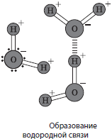
Water(H 2 O) - the most common inorganic substance of the cell, which has unique physicochemical properties. It has no taste, no color, no smell. Density and viscosity of all substances are estimated by water. Like many other substances, water can be in three states of aggregation: solid (ice), liquid and gaseous (steam). The melting point of water is $0°$C, the boiling point is $100°$C, however, the dissolution of other substances in water can change these characteristics. The heat capacity of water is also quite high - 4200 kJ / mol K, which makes it possible for it to take part in the processes of thermoregulation. In a water molecule, hydrogen atoms are located at an angle of $105°$, while the common electron pairs are pulled away by the more electronegative oxygen atom. This determines the dipole properties of water molecules (one of their ends is positively charged and the other negatively) and the possibility of formation of hydrogen bonds between water molecules. The adhesion of water molecules underlies the phenomenon of surface tension, capillarity and the properties of water as a universal solvent. As a result, all substances are divided into soluble in water (hydrophilic) and insoluble in it (hydrophobic). Thanks to these unique properties, it is predetermined that water has become the basis of life on Earth.
The average water content in the cells of the body is not the same and may change with age. So, in a one and a half month old human embryo, the water content in the cells reaches 97.5%, in an eight month old - 83%, in a newborn it decreases to 74%, and in an adult it averages 66%. However, body cells differ in water content. So, the bones contain about 20% water, the liver - 70%, and the brain - 86%. On the whole, it can be said that the concentration of water in cells is directly proportional to the metabolic rate.
mineral salts may be in dissolved or undissolved states. Soluble salts dissociate into ions - cations and anions. The most important cations are potassium and sodium ions, which facilitate the transfer of substances across the membrane and participate in the occurrence and conduction of a nerve impulse; as well as calcium ions, which takes part in the processes of contraction of muscle fibers and blood clotting; magnesium, which is part of chlorophyll; iron, which is part of a number of proteins, including hemoglobin. The most important anions are the phosphate anion, which is part of ATP and nucleic acids, and the carbonic acid residue, which softens fluctuations in the pH of the medium. Ions of mineral salts provide both the penetration of water itself into the cell and its retention in it. If the concentration of salts in the environment is lower than in the cell, then water penetrates into the cell. Also, ions determine the buffer properties of the cytoplasm, i.e., its ability to maintain a constant slightly alkaline pH of the cytoplasm, despite the constant formation of acidic and alkaline products in the cell.
Insoluble salts(CaCO 3, Ca 3 (PO 4) 2, etc.) are part of the bones, teeth, shells and shells of unicellular and multicellular animals.
In addition, other inorganic compounds, such as acids and oxides, can be produced in organisms. So, parietal cells of the human stomach produce hydrochloric acid, which activates the digestive enzyme pepsin, and silicon oxide impregnates the cell walls of horsetails and forms diatom shells. In recent years, the role of nitric oxide (II) in signaling in cells and the body has also been investigated.
organic matter
General characteristics of the organic substances of the cell
The organic substances of a cell can be represented by both relatively simple molecules and more complex ones. In cases where a complex molecule (macromolecule) is formed by a significant number of repeating simpler molecules, it is called polymer, and structural units - monomers. Depending on whether the units of polymers are repeated or not, they are classified as regular or irregular. Polymers make up to 90% of the dry matter mass of the cell. They belong to three main classes of organic compounds - carbohydrates (polysaccharides), proteins and nucleic acids. Regular polymers are polysaccharides, while proteins and nucleic acids are irregular. In proteins and nucleic acids, the sequence of monomers is extremely important, since they perform an informational function.

Carbohydrates
Carbohydrates- these are organic compounds, which mainly include three chemical elements - carbon, hydrogen and oxygen, although a number of carbohydrates also contain nitrogen or sulfur. The general formula for carbohydrates is C m (H 2 O) n. They are divided into simple and complex carbohydrates.
Simple carbohydrates (monosaccharides) contain a single sugar molecule that cannot be broken down into simpler ones. These are crystalline substances, sweet in taste and highly soluble in water. Monosaccharides take an active part in the metabolism in the cell and are part of complex carbohydrates - oligosaccharides and polysaccharides.
Monosaccharides are classified by the number of carbon atoms (C 3 -C 9), for example, pentoses(C 5) and hexoses(From 6). Pentoses include ribose and deoxyribose. Ribose is part of RNA and ATP. Deoxyribose is a component of DNA. Hexoses (C 6 H 12 O 6) are glucose, fructose, galactose, etc. Glucose(grape sugar) is found in all organisms, including human blood, as it is an energy reserve. It is part of many complex sugars: sucrose, lactose, maltose, starch, cellulose, etc. Fructose(fruit sugar) is found in the highest concentrations in fruits, honey, sugar beet root crops. It not only takes an active part in metabolic processes, but also is part of sucrose and some polysaccharides, such as insulin.
Most monosaccharides are able to give a silver mirror reaction and reduce copper by adding Fehling's liquid (a mixture of solutions of copper (II) sulfate and potassium-sodium tartrate) and boiling.

To oligosaccharides include carbohydrates formed by several monosaccharide residues. They are generally also highly soluble in water and are sweet in taste. Depending on the number of these residues, disaccharides (two residues), trisaccharides (three), etc. are distinguished. Disaccharides include sucrose, lactose, maltose, etc. sucrose(beet or cane sugar) consists of residues of glucose and fructose, it is found in the storage organs of some plants. Especially a lot of sucrose in the roots of sugar beet and sugar cane, where they are obtained in an industrial way. It serves as a benchmark for the sweetness of carbohydrates. Lactose, or milk sugar, formed by residues of glucose and galactose, found in mother's and cow's milk. Maltose(malt sugar) consists of two glucose residues. It is formed during the breakdown of polysaccharides in plant seeds and in the human digestive system, and is used in the production of beer.

Polysaccharides are biopolymers whose monomers are mono- or disaccharide residues. Most polysaccharides are insoluble in water and taste unsweetened. These include starch, glycogen, cellulose and chitin. Starch- This is a white powdery substance that is not wetted by water, but forms a suspension when brewed with hot water - a paste. Starch is actually made up of two polymers, the less branched amylose and the more branched amylopectin (Figure 2.9). The monomer of both amylose and amylopectin is glucose. Starch is the main reserve substance of plants, which accumulates in large quantities in seeds, fruits, tubers, rhizomes and other storage organs of plants. A qualitative reaction to starch is a reaction with iodine, in which the starch turns blue-violet.

Glycogen(animal starch) is a reserve polysaccharide of animals and fungi, which in humans accumulates in the largest quantities in the muscles and liver. It is also insoluble in water and tastes unsweetened. The monomer of glycogen is glucose. Compared to starch molecules, glycogen molecules are even more branched.
Cellulose, or cellulose, - the main reference polysaccharide of plants. The monomer of cellulose is glucose. Unbranched cellulose molecules form bundles that are part of the cell walls of plants. Cellulose is the basis of wood, it is used in construction, in the production of textiles, paper, alcohol and many organic substances. Cellulose is chemically inert and does not dissolve in either acids or alkalis. It is also not broken down by the enzymes of the human digestive system, but bacteria in the large intestine help digest it. In addition, fiber stimulates the contraction of the walls of the gastrointestinal tract, helping to improve its work.

Chitin is a polysaccharide, the monomer of which is a nitrogen-containing monosaccharide. It is part of the cell walls of fungi and arthropod shells. In the human digestive system, there is also no enzyme for digesting chitin, only some bacteria have it.
Functions of carbohydrates. Carbohydrates perform plastic (construction), energy, storage and support functions in the cell. They form the cell walls of plants and fungi. The energy value of the breakdown of 1 g of carbohydrates is 17.2 kJ. Glucose, fructose, sucrose, starch and glycogen are reserve substances. Carbohydrates can also be part of complex lipids and proteins, forming glycolipids and glycoproteins, in particular in cell membranes. No less important is the role of carbohydrates in the intercellular recognition and perception of environmental signals, since they act as receptors in the composition of glycoproteins.
Lipids
Lipids is a chemically heterogeneous group of low molecular weight substances with hydrophobic properties. These substances are insoluble in water, form emulsions in it, but are readily soluble in organic solvents. Lipids are oily to the touch, many of them leave characteristic non-drying traces on paper. Together with proteins and carbohydrates, they are one of the main components of cells. The content of lipids in different cells is not the same, especially a lot of them in the seeds and fruits of some plants, in the liver, heart, blood.
Depending on the structure of the molecule, lipids are divided into simple and complex. To simple lipids include neutral lipids (fats), waxes and steroids. Complex lipids also contain another, non-lipid component. The most important of them are phospholipids, glycolipids, etc.
Fats are esters of the trihydric alcohol glycerol and higher fatty acids. Most fatty acids contain 14-22 carbon atoms. Among them there are both saturated and unsaturated, that is, containing double bonds. Of the saturated fatty acids, palmitic and stearic acids are most common, and of the unsaturated fatty acids, oleic. Some unsaturated fatty acids are not synthesized in the human body or are synthesized in insufficient quantities, and therefore are indispensable. Glycerol residues form hydrophilic heads, while fatty acid residues form hydrophobic tails.

Fats perform mainly a storage function in cells and serve as a source of energy. They are rich in subcutaneous fatty tissue, which performs shock-absorbing and thermal insulation functions, and in aquatic animals it also increases buoyancy. Plant fats mostly contain unsaturated fatty acids, as a result of which they are liquid and are called oils. Oils are found in the seeds of many plants, such as sunflower, soybeans, rapeseed, etc.
Waxes are esters and mixtures of fatty acids and fatty alcohols. In plants, they form a film on the surface of the leaf, which protects against evaporation, the penetration of pathogens, etc. In a number of animals, they cover the body or serve to build honeycombs.
To steroids include lipids such as cholesterol, an essential component of cell membranes, as well as sex hormones estradiol, testosterone, vitamin D, etc.
Phospholipids, in addition to residues of glycerol and fatty acids, contain a residue of orthophosphoric acid. They are part of cell membranes and provide their barrier properties.
Glycolipids are also components of membranes, but their content there is low. The non-lipid part of glycolipids are carbohydrates.
Functions of lipids. Lipids perform plastic (building), energy, storage, protective, excretory and regulatory functions in the cell, in addition, they are vitamins. It is an essential component of cell membranes. When splitting 1 g of lipids, 38.9 kJ of energy is released. They are deposited in the reserve in various organs of plants and animals. In addition, subcutaneous fatty tissue protects internal organs from hypothermia or overheating, as well as shock. The regulatory function of lipids is due to the fact that some of them are hormones. The fat body of insects serves for excretion.
Squirrels
Squirrels- These are high-molecular compounds, biopolymers, the monomers of which are amino acids linked by peptide bonds.
amino acid called an organic compound having an amino group, a carboxyl group and a radical. In total, about 200 amino acids are found in nature, which differ in radicals and the mutual arrangement of functional groups, but only 20 of them can be part of proteins. These amino acids are called proteinogenic.

Unfortunately, not all proteinogenic amino acids can be synthesized in the human body, so they are divided into interchangeable and irreplaceable. Non-essential amino acids are formed in the human body in the required amount, and irreplaceable- No. They must come from food, but can also be partially synthesized by intestinal microorganisms. There are 8 fully essential amino acids. These include valine, isoleucine, leucine, lysine, methionine, threonine, tryptophan, and phenylalanine. Despite the fact that absolutely all proteinogenic amino acids are synthesized in plants, vegetable proteins are incomplete because they do not contain a complete set of amino acids, moreover, the presence of protein in the vegetative parts of plants rarely exceeds 1-2% of the mass. Therefore, it is necessary to eat proteins not only of vegetable, but also of animal origin.
A sequence of two amino acids linked by peptide bonds is called dipeptide, out of three tripeptide etc. Among the peptides there are such important compounds as hormones (oxytocin, vasopressin), antibiotics, etc. A chain of more than twenty amino acids is called polypeptide, and polypeptides containing more than 60 amino acid residues are proteins.

Levels of protein structural organization. Proteins can have primary, secondary, tertiary and quaternary structures.
Primary structure of a protein- this is linear amino acid sequence linked by a peptide bond. The primary structure ultimately determines the specificity of the protein and its uniqueness, because even if we assume that the average protein contains 500 amino acid residues, then the number of possible combinations is 20,500. Therefore, a change in the location of at least one amino acid in the primary structure entails a change secondary and higher structures, as well as the properties of the protein as a whole.

Structural features of the protein determine its spatial packing - the emergence of secondary and tertiary structures.
secondary structure is the spatial arrangement of a protein molecule in the form spirals or folds held by hydrogen bonds between the oxygen and hydrogen atoms of peptide groups of different turns of the helix or folds. Many proteins contain more or less long regions with a secondary structure. These are, for example, keratins of hair and nails, silk fibroin.

Tertiary structure squirrel ( globule) is also a form of spatial folding of the polypeptide chain, held by hydrophobic, hydrogen, disulfide (S-S) and other bonds. It is characteristic of most body proteins, such as muscle myoglobin.

Quaternary structure- the most complex, formed by several polypeptide chains connected mainly by the same bonds as in the tertiary (hydrophobic, ionic and hydrogen), as well as other weak interactions. The quaternary structure is characteristic of a few proteins, such as hemoglobin, chlorophyll, etc.

The shape of the molecule is fibrillar and globular proteins. The first of them are elongated, like, for example, connective tissue collagen or hair and nail keratins. Globular proteins are in the form of a ball (globules), like muscle myoglobin.
Simple and complex proteins. Proteins can be simple and complex. Simple proteins are made up of only amino acids, while complex proteins (lipoproteins, chromoproteins, glycoproteins, nucleoproteins, etc.) contain protein and non-protein parts. Chromoproteins contain a colored non-protein portion. These include hemoglobin, myoglobin, chlorophyll, cytochromes, etc. Thus, in the composition of hemoglobin, each of the four polypeptide chains of the globin protein is associated with a non-protein part - heme, in the center of which there is an iron ion, which gives hemoglobin a red color. Non-protein part lipoproteins is a lipid and glycoproteins- carbohydrate. Both lipoproteins and glycoproteins are part of cell membranes. Nucleoproteins are complexes of proteins and nucleic acids (DNA and RNA). They perform the most important functions in the processes of storage and transmission of hereditary information.
Protein properties. Many proteins are highly soluble in water, but there are some among them that dissolve only in solutions of salts, alkalis, acids, or organic solvents. The structure of a protein molecule and its functional activity depend on environmental conditions. The loss of a protein molecule of its structure while maintaining the primary is called denaturation.

Denaturation occurs due to changes in temperature, pH, atmospheric pressure, under the influence of acids, alkalis, salts of heavy metals, organic solvents, etc. The reverse process of restoring secondary and higher structures is called renaturation, however, it is not always possible. The complete breakdown of a protein molecule is called destruction.
Protein functions. Proteins perform a number of functions in the cell: plastic (construction), catalytic (enzymatic), energy, signal (receptor), contractile (motor), transport, protective, regulatory and storage.
The building function of proteins is associated with their presence in cell membranes and structural components of the cell. Energy - due to the fact that during the breakdown of 1 g of protein, 17.2 kJ of energy is released. Membrane receptor proteins are actively involved in the perception of environmental signals and their transmission through the cell, as well as in intercellular recognition. Without proteins, the movement of cells and organisms as a whole is impossible, since they form the basis of flagella and cilia, and also provide muscle contraction and movement of intracellular components. In the blood of humans and many animals, the protein hemoglobin carries oxygen and part of carbon dioxide, while other proteins transport ions and electrons. The protective role of proteins is associated primarily with immunity, since the interferon protein is able to destroy many viruses, and antibody proteins inhibit the development of bacteria and other foreign agents. There are many hormones among proteins and peptides, for example, the pancreatic hormone insulin, which regulates the concentration of glucose in the blood. In some organisms, proteins can be stored in reserve, as in legumes in seeds, or the proteins of a chicken egg.
Nucleic acids
Nucleic acids are biopolymers whose monomers are nucleotides. Currently, two types of nucleic acids are known: ribonucleic (RNA) and deoxyribonucleic (DNA).
Nucleotide formed by a nitrogenous base, a pentose sugar residue, and a phosphoric acid residue. The features of nucleotides are mainly determined by the nitrogenous bases that make up their composition, therefore, even conditionally, nucleotides are designated by the first letters of their names. The composition of nucleotides can include five nitrogenous bases: adenine (A), guanine (G), thymine (T), uracil (U) and cytosine (C). The pentoses of nucleotides - ribose and deoxyribose - determine which nucleotide will be formed - ribonucleotide or deoxyribonucleotide. Ribonucleotides are RNA monomers, they can act as signal molecules (cAMP) and be part of high-energy compounds, such as ATP, and coenzymes, such as NADP, NAD, FAD, etc., and deoxyribonucleotides are part of DNA.

Deoxyribonucleic acid (DNA)- double-stranded biopolymer, the monomers of which are deoxyribonucleotides. The composition of deoxyribonucleotides includes only four nitrogenous bases out of five possible - adenine (A), thymine (T), guanine (G) or cytosine (C), as well as deoxyribose and phosphoric acid residues. Nucleotides in the DNA chain are interconnected through orthophosphoric acid residues, forming a phosphodiester bond. When a double-stranded molecule is formed, the nitrogenous bases are directed inward of the molecule. However, the connection of DNA chains does not occur randomly - the nitrogenous bases of different chains are interconnected by hydrogen bonds according to the principle of complementarity: adenine is connected to thymine by two hydrogen bonds (A \u003d T), and guanine and cytosine by three (G $ ≡ $ C).

For her were set Chargaff rules:
- The number of DNA nucleotides containing adenine is equal to the number of nucleotides containing thymine (A=T).
- The number of DNA nucleotides containing guanine is equal to the number of nucleotides containing cytosine (G$≡$C).
- The sum of deoxyribonucleotides containing adenine and guanine is equal to the sum of deoxyribonucleotides containing thymine and cytosine (A+G = T+C).
- The ratio of the sum of deoxyribonucleotides containing adenine and thymine to the sum of deoxyribonucleotides containing guanine and cytosine depends on the type of organism.
The structure of DNA was deciphered by F. Crick and D. Watson (Nobel Prize in Physiology or Medicine, 1962). According to their model, the DNA molecule is a right-handed double helix. The distance between nucleotides in the DNA chain is 0.34 nm.

The most important property of DNA is the ability to replicate (self-doubling). The main function of DNA is the storage and transmission of hereditary information, which is written in the form of nucleotide sequences. The stability of the DNA molecule is maintained by powerful repair (recovery) systems, but even they are not able to completely eliminate adverse effects, which ultimately leads to mutations. The DNA of eukaryotic cells is concentrated in the nucleus, mitochondria and plastids, while prokaryotic cells are located directly in the cytoplasm. Nuclear DNA is the basis of chromosomes, it is represented by open molecules. DNA of mitochondria, plastids and prokaryotes has a circular shape.
Ribonucleic acid (RNA)- a biopolymer whose monomers are ribonucleotides. They also contain four nitrogenous bases - adenine (A), uracil (U), guanine (G) or cytosine (C), thereby differing from DNA in one of the bases (instead of thymine, RNA contains uracil). The pentose sugar residue in ribonucleotides is represented by ribose. RNA is mostly single-stranded molecules, with the exception of some viral ones. There are three main types of RNA: informational, or template (mRNA, mRNA), ribosomal (rRNA) and transport (tRNA). All of them are formed in the process transcriptions- rewriting from DNA molecules.
and RNAs make up the smallest fraction of RNA in a cell (2-4%), which is offset by their diversity, since one cell can contain thousands of different mRNAs. These are single-stranded molecules that are templates for the synthesis of polypeptide chains. Information about the structure of the protein is recorded in them in the form of sequences of nucleotides, and each amino acid encodes a triplet of nucleotides - codon.
R RNA is the most numerous type of RNA in the cell (up to 80%). Their molecular weight averages 3000-5000; are formed in the nucleoli and are part of the cellular organelles - ribosomes. rRNAs also appear to play a role in protein synthesis.
t RNA is the smallest of the RNA molecules, as it contains only 73-85 nucleotides. Their share of the total amount of cell RNA is about 16%. The function of tRNA is the transport of amino acids to the site of protein synthesis (on ribosomes). The shape of the tRNA molecule resembles a clover leaf. At one end of the molecule there is a site for attaching an amino acid, and in one of the loops there is a triplet of nucleotides that is complementary to the mRNA codon and determines which amino acid the tRNA will carry - anticodon.

All types of RNA take an active part in the implementation of hereditary information, which is rewritten from DNA to mRNA, and on the latter protein synthesis is carried out. tRNA in the process of protein synthesis delivers amino acids to ribosomes, and rRNA is part of the ribosomes directly.
Adenosine triphosphoric acid (ATP) is a nucleotide containing, in addition to the nitrogenous base of adenine and a ribose residue, three phosphoric acid residues. The bonds between the last two phosphorus residues are macroergic (42 kJ / mol of energy is released during splitting), while the standard chemical bond during splitting gives 12 kJ / mol. If energy is needed, the macroergic bond of ATP is split, adenosine diphosphoric acid (ADP), a phosphorus residue are formed, and energy is released:
ATP + H 2 O $→$ ADP + H 3 PO 4 + 42 kJ.
ADP can also be broken down to form AMP (adenosine monophosphoric acid) and a phosphoric acid residue:
ADP + H 2 O $→$ AMP + H 3 PO 4 + 42 kJ.
In the process of energy metabolism (during respiration, fermentation), as well as in the process of photosynthesis, ADP attaches a phosphorus residue and turns into ATP. The ATP recovery reaction is called phosphorylation. ATP is a universal source of energy for all life processes of living organisms.
The study of the chemical composition of the cells of all living organisms has shown that they contain the same chemical elements, chemicals that perform the same functions. Moreover, a piece of DNA transferred from one organism to another will work in it, and a protein synthesized by bacteria or fungi will act as a hormone or enzyme in the human body. This is one of the proofs of the unity of the origin of the organic world.

Cell structure. The relationship of the structure and functions of the parts and organelles of the cell is the basis of its integrity
Cell structure
The structure of prokaryotic and eukaryotic cells
The main structural components of cells are the plasma membrane, cytoplasm and hereditary apparatus. Depending on the characteristics of the organization, two main types of cells are distinguished: prokaryotic and eukaryotic. The main difference between prokaryotic and eukaryotic cells is the organization of their hereditary apparatus: in prokaryotes it is located directly in the cytoplasm (this area of the cytoplasm is called nucleoid) and is not separated from it by membrane structures, while in eukaryotes most of the DNA is concentrated in the nucleus, surrounded by a double membrane. In addition, the genetic information of prokaryotic cells, located in the nucleoid, is recorded in the circular DNA molecule, while in eukaryotes the DNA molecules are not closed.
Unlike eukaryotes, the cytoplasm of prokaryotic cells also contains a small amount of organelles, while eukaryotic cells are characterized by a significant variety of these structures.
The structure and functions of biological membranes
The structure of the biomembrane. The cell-bounding membranes and membrane organelles of eukaryotic cells share a common chemical composition and structure. They include lipids, proteins and carbohydrates. Membrane lipids are mainly represented by phospholipids and cholesterol. Most membrane proteins are complex proteins such as glycoproteins. Carbohydrates do not occur on their own in the membrane, they are associated with proteins and lipids. The thickness of the membranes is 7-10 nm.

According to the currently accepted fluid mosaic model of membrane structure, lipids form a double layer, or lipid bilayer, in which the hydrophilic "heads" of lipid molecules are turned outward, and the hydrophobic "tails" are hidden inside the membrane. These “tails”, due to their hydrophobicity, ensure the separation of the aqueous phases of the internal environment of the cell and its environment. Proteins are associated with lipids through various types of interactions. Some of the proteins are located on the surface of the membrane. Such proteins are called peripheral, or superficial. Other proteins are partially or completely immersed in the membrane - these are integral, or submerged proteins. Membrane proteins perform structural, transport, catalytic, receptor and other functions.
Membranes are not like crystals, their components are constantly in motion, as a result of which gaps appear between lipid molecules - pores through which various substances can enter or leave the cell.
Biological membranes differ in their location in the cell, their chemical composition, and their functions. The main types of membranes are plasma and internal. plasma membrane contains about 45% lipids (including glycolipids), 50% proteins and 5% carbohydrates. Chains of carbohydrates that make up complex proteins-glycoproteins and complex lipids-glycolipids protrude above the surface of the membrane. Plasmalemmal glycoproteins are extremely specific. So, for example, through them there is a mutual recognition of cells, including sperm and eggs.
On the surface of animal cells, carbohydrate chains form a thin surface layer - glycocalyx. It has been found in almost all animal cells, but its severity is not the same (10-50 microns). The glycocalyx provides a direct connection of the cell with the external environment; extracellular digestion occurs in it; receptors are located in the glycocalyx. The cells of bacteria, plants and fungi, in addition to the plasmalemma, are also surrounded by cell membranes.
Internal membranes eukaryotic cells delimit different parts of the cell, forming a kind of "compartments" - compartments, which contributes to the separation of various processes of metabolism and energy. They may differ in chemical composition and functions, but they retain the general plan of the structure.
Membrane functions:
- Limiting. It consists in the fact that they separate the internal space of the cell from the external environment. The membrane is semi-permeable, that is, only those substances that are necessary for the cell can freely overcome it, while there are mechanisms for transporting the necessary substances.
- Receptor. It is associated primarily with the perception of environmental signals and the transfer of this information into the cell. Special receptor proteins are responsible for this function. Membrane proteins are also responsible for cellular recognition according to the "friend or foe" principle, as well as for the formation of intercellular connections, the most studied of which are the synapses of nerve cells.
- catalytic. Numerous enzyme complexes are located on the membranes, as a result of which intensive synthetic processes take place on them.
- Energy transforming. Associated with the formation of energy, its storage in the form of ATP and expenditure.
- Compartmentalization. The membranes also delimit the space inside the cell, thereby separating the initial substances of the reaction and the enzymes that can carry out the corresponding reactions.
- Formation of intercellular contacts. Despite the fact that the thickness of the membrane is so small that it cannot be distinguished with the naked eye, on the one hand, it serves as a fairly reliable barrier for ions and molecules, especially water-soluble ones, and on the other hand, it ensures their transfer into the cell and out.
- Transport.

membrane transport. Due to the fact that cells as elementary biological systems are open systems, to ensure metabolism and energy, maintain homeostasis, growth, irritability and other processes, the transfer of substances through the membrane is required - membrane transport. Currently, the transport of substances across the cell membrane is divided into active, passive, endo- and exocytosis.
Passive transport is a type of transport that occurs without the expenditure of energy from a higher concentration to a lower one. Lipid-soluble small non-polar molecules (O 2, CO 2) easily penetrate the cell by simple diffusion. Insoluble in lipids, including charged small particles, are picked up by carrier proteins or pass through special channels (glucose, amino acids, K +, PO 4 3-). This type of passive transport is called facilitated diffusion. Water enters the cell through pores in the lipid phase, as well as through special channels lined with proteins. The transport of water across a membrane is called osmosis.

Osmosis is extremely important in the life of a cell, because if it is placed in a solution with a higher concentration of salts than in a cell solution, then water will begin to leave the cell, and the volume of living contents will begin to decrease. In animal cells, the cell as a whole shrinks, and in plant cells, the cytoplasm lags behind the cell wall, which is called plasmolysis. When a cell is placed in a solution less concentrated than the cytoplasm, water is transported in the opposite direction - into the cell. However, there are limits to the extensibility of the cytoplasmic membrane, and the animal cell eventually ruptures, while in the plant cell this is not allowed by a strong cell wall. The phenomenon of filling the entire internal space of the cell with cellular contents is called deplasmolysis. The intracellular salt concentration should be taken into account when preparing drugs, especially for intravenous administration, as this can lead to damage to blood cells (for this, a saline solution with a concentration of 0.9% sodium chloride is used). This is no less important in the cultivation of cells and tissues, as well as organs of animals and plants.

active transport proceeds with the expenditure of ATP energy from a lower concentration of a substance to a higher one. It is carried out with the help of special proteins-pumps. Proteins pump ions K +, Na +, Ca 2+ and others through the membrane, which contributes to the transport of the most important organic substances, as well as the emergence of nerve impulses, etc.
Endocytosis- this is an active process of absorption of substances by the cell, in which the membrane forms invaginations, and then forms membrane vesicles - phagosomes, which contain absorbed objects. The primary lysosome then fuses with the phagosome to form secondary lysosome, or phagolysosome, or digestive vacuole. The contents of the vesicle are cleaved by lysosome enzymes, and the cleavage products are absorbed and assimilated by the cell. Undigested residues are removed from the cell by exocytosis. There are two main types of endocytosis: phagocytosis and pinocytosis.
Phagocytosis is the process of capture by the cell surface and absorption of solid particles by the cell, and pinocytosis- liquids. Phagocytosis occurs mainly in animal cells (single-celled animals, human leukocytes), it provides their nutrition, and often the protection of the body. By way of pinocytosis, the absorption of proteins, antigen-antibody complexes in the process of immune reactions, etc. occurs. However, many viruses also enter the cell by way of pinocytosis or phagocytosis. In the cells of plants and fungi, phagocytosis is practically impossible, since they are surrounded by strong cell membranes.

Exocytosis is the reverse process of endocytosis. Thus, undigested food residues are released from the digestive vacuoles, the substances necessary for the life of the cell and the organism as a whole are removed. For example, the transmission of nerve impulses occurs due to the release of chemical messengers by the neuron that sends the impulse - mediators, and in plant cells, auxiliary carbohydrates of the cell membrane are released in this way.
Cell walls of plant cells, fungi and bacteria. Outside of the membrane, the cell can secrete a strong framework - cell membrane, or cell wall.
In plants, the cell wall is made up of cellulose packed in bundles of 50-100 molecules. The gaps between them are filled with water and other carbohydrates. The plant cell membrane is pierced by tubules - plasmodesmata through which the membranes of the endoplasmic reticulum pass. The plasmodesmata transport substances between cells. However, the transport of substances, such as water, can also occur along the cell walls themselves. Over time, various substances, including tannins or fat-like substances, accumulate in the cell membrane of plants, which leads to lignification or corking of the cell wall itself, the displacement of water and the death of cellular contents. Between the cell walls of neighboring plant cells there are jelly-like pads - middle plates that fasten them together and cement the plant body as a whole. They are destroyed only in the process of fruit ripening and when the leaves fall.

The cell walls of fungal cells are formed chitin- a carbohydrate containing nitrogen. They are strong enough and are the outer skeleton of the cell, but still, like in plants, they prevent phagocytosis.
In bacteria, the cell wall contains a carbohydrate with fragments of peptides - murein, however, its content varies significantly in different groups of bacteria. On top of the cell wall, other polysaccharides can also be released, forming a mucous capsule that protects bacteria from external influences.
The shell determines the shape of the cell, serves as a mechanical support, performs a protective function, provides the osmotic properties of the cell, limiting the stretching of the living contents and preventing the rupture of the cell, which increases due to the influx of water. In addition, water and substances dissolved in it overcome the cell wall before entering the cytoplasm or, conversely, when leaving it, while water is transported along the cell walls faster than through the cytoplasm.
Cytoplasm
Cytoplasm is the interior of the cell. All organelles of the cell, the nucleus and various waste products are immersed in it.
The cytoplasm connects all parts of the cell with each other, numerous metabolic reactions take place in it. The cytoplasm is separated from the environment and divided into compartments by membranes, that is, cells have a membrane structure. It can be in two states - sol and gel. Sol- this is a semi-liquid, jelly-like state of the cytoplasm, in which vital processes proceed most intensively, and gel- a denser, gelatinous state that impedes the flow of chemical reactions and the transport of substances.
The liquid part of the cytoplasm without organelles is called hyaloplasm. Hyaloplasm, or cytosol, is a colloidal solution in which there is a kind of suspension of fairly large particles, such as proteins, surrounded by dipoles of water molecules. The sedimentation of this suspension does not occur due to the fact that they have the same charge and repel each other.
Organelles
Organelles- These are permanent components of the cell that perform certain functions.
Depending on the structural features, they are divided into membrane and non-membrane. Membrane organelles, in turn, are referred to as single-membrane (endoplasmic reticulum, Golgi complex and lysosomes) or double-membrane (mitochondria, plastids and nucleus). Non-membrane organelles are ribosomes, microtubules, microfilaments and the cell center. Of the listed organelles, only ribosomes are inherent in prokaryotes.
The structure and functions of the nucleus. Nucleus- a large two-membrane organelle lying in the center of the cell or on its periphery. The size of the nucleus can vary within 3-35 microns. The shape of the nucleus is more often spherical or ellipsoid, but there are also rod-shaped, spindle-shaped, bean-shaped, lobed and even segmented nuclei. Some researchers believe that the shape of the nucleus corresponds to the shape of the cell itself.
Most cells have one nucleus, but, for example, in liver and heart cells there can be two, and in a number of neurons - up to 15. Skeletal muscle fibers usually contain many nuclei, but they are not cells in the full sense of the word, since they are formed in the result of the fusion of several cells.
The core is surrounded nuclear envelope, and its interior space is filled nuclear juice, or nucleoplasm (karyoplasm) in which are immersed chromatin and nucleolus. The nucleus performs such important functions as the storage and transmission of hereditary information, as well as the control of cell vital activity.

The role of the nucleus in the transmission of hereditary information has been convincingly proven in experiments with the green algae acetabularia. In a single giant cell, reaching a length of 5 cm, a hat, a leg and a rhizoid are distinguished. Moreover, it contains only one nucleus located in the rhizoid. In the 1930s, I. Hemmerling transplanted the nucleus of one species of acetabularia with a green color into a rhizoid of another species, with a brown color, in which the nucleus was removed. After some time, the plant with the transplanted nucleus grew a new cap, like the algae-donor of the nucleus. At the same time, the cap or stalk separated from the rhizoid, which did not contain a nucleus, died after some time.

nuclear envelope It is formed by two membranes - outer and inner, between which there is a space. The intermembrane space communicates with the cavity of the rough endoplasmic reticulum, and the outer membrane of the nucleus can carry ribosomes. The nuclear envelope is permeated with numerous pores, edged with special proteins. Substances are transported through the pores: the necessary proteins (including enzymes), ions, nucleotides and other substances enter the nucleus, and RNA molecules, waste proteins, subunits of ribosomes leave it. Thus, the functions of the nuclear envelope are the separation of the contents of the nucleus from the cytoplasm, as well as the regulation of the metabolism between the nucleus and the cytoplasm.
Nucleoplasm called the contents of the nucleus, in which the chromatin and nucleolus are immersed. It is a colloidal solution, chemically reminiscent of the cytoplasm. Enzymes of the nucleoplasm catalyze the exchange of amino acids, nucleotides, proteins, etc. The nucleoplasm is connected to the hyaloplasm through nuclear pores. The functions of the nucleoplasm, like the hyaloplasm, are to ensure the interconnection of all structural components of the nucleus and the implementation of a number of enzymatic reactions.
chromatin called a set of thin threads and granules immersed in the nucleoplasm. It can only be detected by staining, since the refractive indices of chromatin and nucleoplasm are approximately the same. The filamentous component of chromatin is called euchromatin, and granular heterochromatin. Euchromatin is weakly compacted, since hereditary information is read from it, while more spiralized heterochromatin is genetically inactive.
Chromatin is a structural modification of chromosomes in a non-dividing nucleus. Thus, chromosomes are constantly present in the nucleus; only their state changes depending on the function that the nucleus performs at the moment.
Chromatin mainly consists of nucleoproteins (deoxyribonucleoproteins and ribonucleoproteins), as well as enzymes, the most important of which are associated with the synthesis of nucleic acids, and some other substances.
The functions of chromatin consist, firstly, in the synthesis of nucleic acids specific for a given organism, which direct the synthesis of specific proteins, and secondly, in the transfer of hereditary properties from the mother cell to daughter cells, for which chromatin threads are packed into chromosomes during division.
nucleolus- a spherical body, clearly visible under a microscope with a diameter of 1-3 microns. It is formed in chromatin regions that encode information about the structure of rRNA and ribosome proteins. The nucleolus in the nucleus is often one, but in those cells where intensive vital processes take place, there may be two or more nucleoli. The functions of the nucleoli are the synthesis of rRNA and the assembly of ribosome subunits by combining rRNA with proteins coming from the cytoplasm.
Mitochondria- two-membrane organelles of a round, oval or rod-shaped shape, although spiral-shaped ones are also found (in spermatozoa). Mitochondria are up to 1 µm in diameter and up to 7 µm in length. The space inside the mitochondria is filled with matrix. Matrix It is the main substance of mitochondria. A circular DNA molecule and ribosomes are immersed in it. The outer membrane of mitochondria is smooth and impermeable to many substances. The inner membrane has outgrowths - cristae, which increase the surface area of membranes for chemical reactions to occur. On the surface of the membrane are numerous protein complexes that make up the so-called respiratory chain, as well as mushroom-shaped enzymes of ATP synthetase. In mitochondria, the aerobic stage of respiration takes place, during which ATP is synthesized.

plastids- large two-membrane organelles, characteristic only for plant cells. The inner space of plastids is filled stroma, or matrix. In the stroma there is a more or less developed system of membrane vesicles - thylakoids, which are collected in piles - grains, as well as its own circular DNA molecule and ribosomes. There are four main types of plastids: chloroplasts, chromoplasts, leucoplasts, and proplastids.
Chloroplasts- These are green plastids with a diameter of 3-10 microns, clearly visible under a microscope. They are found only in the green parts of plants - leaves, young stems, flowers and fruits. Chloroplasts are mostly oval or ellipsoid in shape, but can also be cup-shaped, spiral-shaped, and even lobed. The number of chloroplasts in a cell averages from 10 to 100 pieces. However, for example, in some algae it may be one, have a significant size and complex shape - then it is called chromatophore. In other cases, the number of chloroplasts can reach several hundred, while their size is small. The color of chloroplasts is due to the main pigment of photosynthesis - chlorophyll, although they contain additional pigments - carotenoids. Carotenoids become noticeable only in autumn, when the chlorophyll in aging leaves is destroyed. The main function of chloroplasts is photosynthesis. Light reactions of photosynthesis occur on thylakoid membranes, on which chlorophyll molecules are fixed, and dark reactions occur in the stroma, which contains numerous enzymes.

Chromoplasts are yellow, orange and red plastids containing carotenoid pigments. The shape of chromoplasts can also vary significantly: they are tubular, spherical, crystalline, etc. Chromoplasts give color to flowers and fruits of plants, attracting pollinators and dispersers of seeds and fruits.
Leucoplasts- These are white or colorless plastids, mostly round or oval in shape. They are common in non-photosynthetic parts of plants, such as leaf skins, potato tubers, etc. They store nutrients, most often starch, but in some plants it can be proteins or oil.
Plastids are formed in plant cells from proplastids, which are already present in the cells of the educational tissue and are small two-membrane bodies. At the early stages of development, different types of plastids are able to turn into each other: when exposed to light, the leukoplasts of a potato tuber and the chromoplasts of a carrot root turn green.
Plastids and mitochondria are called semi-autonomous cell organelles, since they have their own DNA molecules and ribosomes, carry out protein synthesis and divide independently of cell division. These features are explained by the origin from unicellular prokaryotic organisms. However, the "independence" of mitochondria and plastids is limited, since their DNA contains too few genes for free existence, while the rest of the information is encoded in the chromosomes of the nucleus, which allows it to control these organelles.
Endoplasmic reticulum (ER), or endoplasmic reticulum (ER), is a single-membrane organelle, which is a network of membrane cavities and tubules, occupying up to 30% of the contents of the cytoplasm. The diameter of ER tubules is about 25–30 nm. There are two types of EPS - rough and smooth. Rough XPS carries ribosomes and is where proteins are synthesized. Smooth EPS devoid of ribosomes. Its function is the synthesis of lipids and carbohydrates, as well as the transport, storage and disposal of toxic substances. It is especially developed in those cells where intensive metabolic processes take place, for example, in liver cells - hepatocytes - and skeletal muscle fibers. Substances synthesized in the EPS are transported to the Golgi apparatus. In the ER, cell membranes are also assembled, but their formation is completed in the Golgi apparatus.
golgi apparatus, or golgi complex, is a single-membrane organelle formed by a system of flat cisterns, tubules and vesicles laced off from them. The structural unit of the Golgi apparatus is dictyosome- a stack of tanks, to one pole of which substances from the ER come, and from the opposite pole, having undergone certain transformations, they are packed into bubbles and sent to other parts of the cell. The diameter of tanks is about 2 microns, and small bubbles are about 20-30 microns. The main functions of the Golgi complex are the synthesis of certain substances and the modification (change) of proteins, lipids and carbohydrates coming from the ER, the final formation of membranes, as well as the transport of substances through the cell, the renewal of its structures and the formation of lysosomes. The Golgi apparatus got its name in honor of the Italian scientist Camillo Golgi, who first discovered this organoid (1898).

Lysosomes- small single-membrane organelles up to 1 micron in diameter, which contain hydrolytic enzymes involved in intracellular digestion. The membranes of lysosomes are poorly permeable for these enzymes, so the performance of their functions by lysosomes is very accurate and targeted. So, they take an active part in the process of phagocytosis, forming digestive vacuoles, and in case of starvation or damage to certain parts of the cell, they digest them without affecting others. Recently, the role of lysosomes in cell death processes has been discovered.
Vacuole- a cavity in the cytoplasm of plant and animal cells, bounded by a membrane and filled with liquid. Digestive and contractile vacuoles are found in protozoan cells. The former take part in the process of phagocytosis, as they break down nutrients. The latter ensure the maintenance of water-salt balance due to osmoregulation. In multicellular animals, digestive vacuoles are mainly found.
In plant cells, vacuoles are always present, they are surrounded by a special membrane and filled with cell sap. The membrane surrounding the vacuole is similar in chemical composition, structure and functions to the plasma membrane. cell sap represents an aqueous solution of various inorganic and organic substances, including mineral salts, organic acids, carbohydrates, proteins, glycosides, alkaloids, etc. The vacuole can occupy up to 90% of the cell volume and push the nucleus to the periphery. This part of the cell performs storage, excretory, osmotic, protective, lysosomal and other functions, since it accumulates nutrients and waste products, it provides water supply and maintains the shape and volume of the cell, and also contains enzymes for the breakdown of many cell components. In addition, the biologically active substances of vacuoles can prevent many animals from eating these plants. In a number of plants, due to the swelling of vacuoles, cell growth occurs by stretching.
Vacuoles are also present in the cells of some fungi and bacteria, but in fungi they perform only the function of osmoregulation, while in cyanobacteria they maintain buoyancy and participate in the processes of nitrogen assimilation from the air.
Ribosomes- small non-membrane organelles with a diameter of 15-20 microns, consisting of two subunits - large and small. Eukaryotic ribosome subunits are assembled in the nucleolus and then transported to the cytoplasm. The ribosomes of prokaryotes, mitochondria, and plastids are smaller than those of eukaryotes. Ribosome subunits include rRNA and proteins.
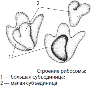
The number of ribosomes in a cell can reach several tens of millions: in the cytoplasm, mitochondria and plastids they are in a free state, and on the rough ER they are in a bound state. They take part in protein synthesis, in particular, they carry out the process of translation - the biosynthesis of a polypeptide chain on an mRNA molecule. On free ribosomes, proteins of hyaloplasm, mitochondria, plastids and own proteins of ribosomes are synthesized, while on ribosomes attached to the rough ER, proteins are translated for excretion from cells, assembly of membranes, formation of lysosomes and vacuoles.
Ribosomes can be located in the hyaloplasm singly or assembled in groups with simultaneous synthesis of several polypeptide chains on one mRNA. These groups of ribosomes are called polyribosomes, or polysomes.

microtubules- These are cylindrical hollow non-membrane organelles that penetrate the entire cytoplasm of the cell. Their diameter is about 25 nm, the wall thickness is 6-8 nm. They are made up of numerous protein molecules. tubulin, which first form 13 strands resembling beads and then assemble into a microtubule. Microtubules form a cytoplasmic reticulum that gives the cell shape and volume, connects the plasma membrane with other parts of the cell, provides transport of substances through the cell, takes part in the movement of the cell and intracellular components, as well as in the division of genetic material. They are part of the cell center and organelles of movement - flagella and cilia.
microfilaments, or microfilaments, are also non-membrane organelles, however, they have a filamentous shape and are formed not by tubulin, but actinome. They take part in the processes of membrane transport, intercellular recognition, division of the cell cytoplasm and in its movement. In muscle cells, the interaction of actin microfilaments with myosin filaments provides contraction.
Microtubules and microfilaments form the inner skeleton of the cell cytoskeleton. It is a complex network of fibers that provide mechanical support for the plasma membrane, determines the shape of the cell, the location of cellular organelles and their movement during cell division.

Cell Center- non-membrane organelle located in animal cells near the nucleus; it is absent in plant cells. Its length is about 0.2–0.3 µm, and its diameter is 0.1–0.15 µm. The cell center is made up of two centrioles lying in mutually perpendicular planes, and radiant sphere from microtubules. Each centriole is formed by nine groups of microtubules, collected in threes, i.e. triplets. The cell center takes part in the assembly of microtubules, the division of the hereditary material of the cell, as well as in the formation of flagella and cilia.

Organelles of movement. Flagella and cilia are outgrowths of cells covered with plasmalemma. These organelles are based on nine pairs of microtubules located along the periphery and two free microtubules in the center. Microtubules are interconnected by various proteins that ensure their coordinated deviation from the axis - oscillation. Fluctuations are energy-dependent, that is, the energy of macroergic bonds of ATP is spent on this process. Restoration of lost flagella and cilia is a function basal bodies, or kinetosomes located at their base.
The length of the cilia is about 10-15 nm, and the length of the flagella is 20-50 microns. Due to the strictly directed movements of the flagella and cilia, not only the movement of unicellular animals, spermatozoa, etc. is carried out, but also the airways are cleared, the egg moves through the fallopian tubes, since all these parts of the human body are lined with ciliated epithelium.

Inclusions
Inclusions- These are non-permanent components of the cell, which are formed and disappear in the course of its life. These include both reserve substances, for example, grains of starch or protein in plant cells, glycogen granules in animal and fungal cells, volutin in bacteria, fat drops in all cell types, and waste products, in particular, undigested food residues as a result of phagocytosis. , forming the so-called residual bodies.
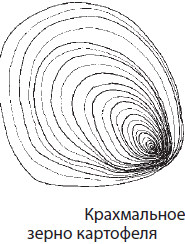
The relationship of the structure and functions of the parts and organelles of the cell is the basis of its integrity
Each of the parts of the cell, on the one hand, is a separate structure with a specific structure and functions, and on the other hand, a component of a more complex system called a cell. Most of the hereditary information of a eukaryotic cell is concentrated in the nucleus, but the nucleus itself is not able to ensure its implementation, since this requires at least the cytoplasm, which acts as the main substance, and ribosomes, on which this synthesis occurs. Most ribosomes are located on the granular endoplasmic reticulum, from where proteins are most often transported to the Golgi complex, and then, after modification, to those parts of the cell for which they are intended, or are excreted. Membrane packaging of proteins and carbohydrates can be integrated into organoid membranes and the cytoplasmic membrane, ensuring their constant renewal. Lysosomes and vacuoles, which perform the most important functions, are also laced from the Golgi complex. For example, without lysosomes, cells would quickly turn into a kind of dump of waste molecules and structures.
All of these processes require energy produced by mitochondria and, in plants, also by chloroplasts. And although these organelles are relatively autonomous, since they have their own DNA molecules, some of their proteins are still encoded by the nuclear genome and synthesized in the cytoplasm.
Thus, the cell is an inseparable unity of its constituent components, each of which performs its own unique function.
Metabolism and energy conversion are properties of living organisms. Energy and plastic metabolism, their relationship. Stages of energy metabolism. Fermentation and respiration. Photosynthesis, its significance, cosmic role. Phases of photosynthesis. Light and dark reactions of photosynthesis, their relationship. Chemosynthesis. The role of chemosynthetic bacteria on Earth
Metabolism and energy conversion - properties of living organisms
The cell can be likened to a miniature chemical factory where hundreds and thousands of chemical reactions take place.
Metabolism- a set of chemical transformations aimed at the preservation and self-reproduction of biological systems.
It includes the intake of substances into the body during nutrition and respiration, intracellular metabolism, or metabolism, as well as the allocation of end products of metabolism.
Metabolism is inextricably linked with the processes of converting one type of energy into another. For example, in the process of photosynthesis, light energy is stored in the form of the energy of chemical bonds of complex organic molecules, and in the process of respiration it is released and spent on the synthesis of new molecules, mechanical and osmotic work, is dissipated in the form of heat, etc.
The flow of chemical reactions in living organisms is ensured by biological catalysts of protein nature - enzymes, or enzymes. Like other catalysts, enzymes accelerate the flow of chemical reactions in the cell by tens and hundreds of thousands of times, and sometimes even make them possible, but do not change either the nature or properties of the final product (products) of the reaction and do not change themselves. Enzymes can be both simple and complex proteins, which, in addition to the protein part, also include a non-protein part - cofactor (coenzyme). Examples of enzymes are salivary amylase, which breaks down polysaccharides during prolonged chewing, and pepsin, which ensures the digestion of proteins in the stomach.
Enzymes differ from non-protein catalysts in their high specificity of action, a significant increase in the reaction rate with their help, as well as the ability to regulate the action by changing the reaction conditions or interacting with various substances. In addition, the conditions under which enzymatic catalysis proceeds differ significantly from those under which non-enzymatic catalysis occurs: the temperature of $37°C$ is optimal for the functioning of enzymes in the human body, the pressure should be close to atmospheric, and the $pH$ of the medium can significantly hesitate. So, for amylase, an alkaline environment is necessary, and for pepsin, an acidic one.
The mechanism of action of enzymes is to reduce the activation energy of substances (substrates) that enter into the reaction due to the formation of intermediate enzyme-substrate complexes.

Energy and plastic metabolism, their relationship
Metabolism consists of two processes simultaneously occurring in the cell: plastic and energy exchanges.
Plastic metabolism (anabolism, assimilation) is a set of synthesis reactions that go with the expenditure of ATP energy. In the process of plastic metabolism, organic substances necessary for the cell are synthesized. Examples of plastic exchange reactions are photosynthesis, protein biosynthesis, and DNA replication (self-doubling).
Energy metabolism (catabolism, dissimilation) is a set of reactions that break down complex substances into simpler ones. As a result of energy metabolism, energy is released, stored in the form of ATP. The most important processes of energy metabolism are respiration and fermentation.
Plastic and energy exchanges are inextricably linked, since organic substances are synthesized in the process of plastic exchange and this requires ATP energy, and in the process of energy metabolism, organic substances are split and energy is released, which will then be spent on synthesis processes.
Organisms receive energy in the process of nutrition, and release it and convert it into an accessible form mainly in the process of respiration. According to the way of nutrition, all organisms are divided into autotrophs and heterotrophs. Autotrophs able to independently synthesize organic substances from inorganic, and heterotrophs use exclusively ready-made organic substances.
Stages of energy metabolism
Despite the complexity of energy metabolism reactions, it is conditionally divided into three stages: preparatory, anaerobic (oxygen-free) and aerobic (oxygen).
On the preparatory stage molecules of polysaccharides, lipids, proteins, nucleic acids break down into simpler ones, for example, glucose, glycerol and fatty acids, amino acids, nucleotides, etc. This stage can take place directly in the cells or in the intestine, from where the split substances are delivered with blood flow.
anaerobic stage energy metabolism is accompanied by further splitting of the monomers of organic compounds to even simpler intermediate products, for example, pyruvic acid, or pyruvate. It does not require the presence of oxygen, and for many organisms living in the silt of swamps or in the human intestine, it is the only way to obtain energy. The anaerobic stage of energy metabolism takes place in the cytoplasm.
Various substances can undergo oxygen-free cleavage, but glucose is often the substrate of the reactions. The process of its oxygen-free splitting is called glycolysis. During glycolysis, the glucose molecule loses four hydrogen atoms, i.e., it is oxidized, and two molecules of pyruvic acid, two ATP molecules and two molecules of the reduced hydrogen carrier $NADH + H^(+)$ are formed:
$C_6H_(12)O_6 + 2H_3PO_4 + 2ADP + 2NAD → 2C_3H_4O_3 + 2ATP + 2NADH + H^(+) + 2H_2O$.
The formation of ATP from ADP occurs due to the direct transfer of a phosphate anion from a previously phosphorylated sugar and is called substrate phosphorylation.
Aerobic Stage energy exchange can occur only in the presence of oxygen, while the intermediate compounds formed in the process of oxygen-free splitting are oxidized to final products (carbon dioxide and water) and most of the energy stored in the chemical bonds of organic compounds is released. It passes into the energy of macroergic bonds of 36 ATP molecules. This stage is also called tissue respiration. In the absence of oxygen, intermediate compounds are converted into other organic substances, a process called fermentation.
Breath
The mechanism of cellular respiration is schematically shown in fig.

Aerobic respiration occurs in mitochondria, while pyruvic acid first loses one carbon atom, which is accompanied by the synthesis of one reducing equivalent of $NADH + H^(+)$ and an acetyl coenzyme A (acetyl-CoA) molecule:
$C_3H_4O_3 + NAD + H~CoA → CH_3CO~CoA + NADH + H^(+) + CO_2$.
Acetyl-CoA in the mitochondrial matrix is involved in a chain of chemical reactions, the totality of which is called Krebs cycle (tricarboxylic acid cycle, citric acid cycle). During these transformations, two ATP molecules are formed, acetyl-CoA is completely oxidized to carbon dioxide, and its hydrogen ions and electrons are attached to the hydrogen carriers $NADH + H^(+)$ and $FADH_2$. Carriers transport hydrogen protons and electrons to the inner membranes of mitochondria, which form cristae. With the help of carrier proteins, hydrogen protons are injected into the intermembrane space, and electrons are transferred along the so-called respiratory chain of enzymes located on the inner membrane of mitochondria and are dumped onto oxygen atoms:
$O_2+2e^(-)→O_2^-$.
It should be noted that some proteins of the respiratory chain contain iron and sulfur.
From the intermembrane space, hydrogen protons are transported back to the mitochondrial matrix with the help of special enzymes - ATP synthases, and the energy released in this case is spent on the synthesis of 34 ATP molecules from each glucose molecule. This process is called oxidative phosphorylation. In the mitochondrial matrix, hydrogen protons react with oxygen radicals to form water:
$4H^(+)+O_2^-→2H_2O$.
The set of reactions of oxygen respiration can be expressed as follows:
$2C_3H_4O_3 + 6O_2 + 36H_3PO_4 + 36ADP → 6CO_2 + 38H_2O + 36ATP.$
The overall breathing equation looks like this:
$C_6H_(12)O_6 + 6O_2 + 38H_3PO_4 + 38ADP → 6CO_2 + 40H_2O + 38ATP.$
Fermentation
In the absence of oxygen or its deficiency, fermentation occurs. Fermentation is an evolutionarily earlier way of obtaining energy than respiration, but it is energetically less profitable, since fermentation produces organic substances that are still rich in energy. There are several main types of fermentation: lactic acid, alcohol, acetic acid, etc. So, in skeletal muscles, in the absence of oxygen during fermentation, pyruvic acid is reduced to lactic acid, while the previously formed reducing equivalents are consumed, and only two ATP molecules remain:
$2C_3H_4O_3 + 2NADH + H^(+) → 2C_3H_6O_3 + 2NAD$.
During fermentation with the help of yeast fungi, pyruvic acid in the presence of oxygen turns into ethyl alcohol and carbon monoxide (IV):
$C_3H_4O_3 + NADH + H^(+) → C_2H_5OH + CO_2 + NAD^(+)$.
During fermentation with the help of microorganisms, pyruvic acid can also form acetic, butyric, formic acids, etc.
ATP obtained as a result of energy metabolism is consumed in the cell for various types of work: chemical, osmotic, electrical, mechanical and regulatory. Chemical work consists in the biosynthesis of proteins, lipids, carbohydrates, nucleic acids and other vital compounds. Osmotic work includes the processes of absorption by the cell and removal from it of substances that are in the extracellular space in concentrations greater than in the cell itself. Electrical work is closely related to osmotic work, since it is as a result of the movement of charged particles through the membranes that the charge of the membrane is formed and the properties of excitability and conductivity are acquired. Mechanical work is associated with the movement of substances and structures inside the cell, as well as the cell as a whole. Regulatory work includes all processes aimed at coordinating processes in the cell.
Photosynthesis, its significance, cosmic role
photosynthesis called the process of converting light energy into the energy of chemical bonds of organic compounds with the participation of chlorophyll.
As a result of photosynthesis, about 150 billion tons of organic matter and approximately 200 billion tons of oxygen are produced annually. This process ensures the circulation of carbon in the biosphere, preventing the accumulation of carbon dioxide and thereby preventing the occurrence of the greenhouse effect and overheating of the Earth. The organic substances formed as a result of photosynthesis are not completely consumed by other organisms, a significant part of them formed mineral deposits (hard and brown coal, oil) over millions of years. Recently, rapeseed oil (“biodiesel”) and alcohol obtained from plant residues have also been used as fuel. From oxygen, under the action of electrical discharges, ozone is formed, which forms an ozone shield that protects all life on Earth from the harmful effects of ultraviolet rays.
Our compatriot, the outstanding plant physiologist K. A. Timiryazev (1843-1920) called the role of photosynthesis “cosmic”, since it connects the Earth with the Sun (space), providing an influx of energy to the planet.
Phases of photosynthesis. Light and dark reactions of photosynthesis, their relationship
In 1905, the English plant physiologist F. Blackman discovered that the rate of photosynthesis cannot increase indefinitely, some factor limits it. Based on this, he proposed the existence of two phases of photosynthesis: light and dark. At low light intensity, the speed of light reactions increases in proportion to the increase in light intensity, and, in addition, these reactions do not depend on temperature, since they do not require enzymes to proceed. Light reactions occur on thylakoid membranes.
The rate of dark reactions, on the contrary, increases with increasing temperature; however, upon reaching the temperature threshold of $30°C$, this growth stops, which indicates the enzymatic nature of these transformations occurring in the stroma. It should be noted that light also has a certain effect on dark reactions, despite the fact that they are called dark.

The light phase of photosynthesis proceeds on thylakoid membranes, which carry several types of protein complexes, the main of which are photosystems I and II, as well as ATP synthase. The composition of photosystems includes pigment complexes, in which, in addition to chlorophyll, there are also carotenoids. Carotenoids trap light in those regions of the spectrum in which chlorophyll does not, and also protect chlorophyll from destruction by high-intensity light.
In addition to pigment complexes, photosystems also include a number of electron acceptor proteins that successively transfer electrons from chlorophyll molecules to each other. The sequence of these proteins is called chloroplast electron transport chain.
A special complex of proteins is also associated with photosystem II, which ensures the release of oxygen during photosynthesis. This oxygen-evolving complex contains manganese and chlorine ions.
AT light phase light quanta, or photons, falling on chlorophyll molecules located on thylakoid membranes, transfer them to an excited state characterized by a higher electron energy. At the same time, excited electrons from the chlorophyll of photosystem I are transferred through a chain of intermediaries to the hydrogen carrier NADP, which adds hydrogen protons, which are always present in an aqueous solution:
$NADP + 2e^(-) + 2H^(+) → NADPH + H^(+)$.
The reduced $NADPH + H^(+)$ will subsequently be used in the dark stage. Electrons from the chlorophyll of photosystem II are also transferred along the electron transport chain, but they fill the “electron holes” in the chlorophyll of photosystem I. The lack of electrons in the chlorophyll of photosystem II is filled by taking away water molecules from water molecules, which occurs with the participation of the oxygen-releasing complex already mentioned above. As a result of the decomposition of water molecules, which is called photolysis, hydrogen protons are formed and molecular oxygen is released, which is a by-product of photosynthesis:
$H_2O → 2H^(+) + 2e^(-) + (1)/(2)O_2$.
Genetic information in a cell. Genes, genetic code and its properties. Matrix nature of biosynthetic reactions. Biosynthesis of protein and nucleic acids
Genetic information in a cell
Reproduction of one's own kind is one of the fundamental properties of the living. Due to this phenomenon, there is a similarity not only between organisms, but also between individual cells, as well as their organelles (mitochondria and plastids). The material basis of this similarity is the transmission of genetic information encrypted in the DNA nucleotide sequence, which is carried out due to the processes of DNA replication (self-doubling). All features and properties of cells and organisms are realized thanks to proteins, the structure of which is primarily determined by the DNA nucleotide sequences. Therefore, it is the biosynthesis of nucleic acids and proteins that is of paramount importance in metabolic processes. The structural unit of hereditary information is the gene.
Genes, genetic code and its properties
Hereditary information in a cell is not monolithic, it is divided into separate "words" - genes.
Gene is the basic unit of genetic information.
The work on the "Human Genome" program, which was carried out simultaneously in several countries and was completed at the beginning of this century, gave us an understanding that a person has only about 25-30 thousand genes, but information from most of our DNA is never read, since it contains a huge number of meaningless sections, repeats and genes encoding features that have lost their meaning for humans (tail, body hair, etc.). In addition, a number of genes responsible for the development of hereditary diseases, as well as drug target genes, have been deciphered. However, the practical application of the results obtained during the implementation of this program is postponed until the genomes of more people are decoded and it becomes clear how they differ.
Genes encoding the primary structure of a protein, ribosomal or transfer RNA are called structural, and genes that provide activation or suppression of reading information from structural genes - regulatory. However, even structural genes contain regulatory regions.
The hereditary information of organisms is encrypted in DNA in the form of certain combinations of nucleotides and their sequence - genetic code. Its properties are: triplet, specificity, universality, redundancy and non-overlapping. In addition, there are no punctuation marks in the genetic code.
Each amino acid is encoded in DNA by three nucleotides. triplet for example, methionine is encoded by the TAC triplet, that is, the triplet code. On the other hand, each triplet encodes only one amino acid, which is its specificity or unambiguity. The genetic code is universal for all living organisms, that is, hereditary information about human proteins can be read by bacteria and vice versa. This testifies to the unity of the origin of the organic world. However, 64 combinations of three nucleotides correspond to only 20 amino acids, as a result of which 2-6 triplets can encode one amino acid, that is, the genetic code is redundant, or degenerate. Three triplets do not have corresponding amino acids, they are called stop codons, as they mark the end of the synthesis of the polypeptide chain.
The sequence of bases in DNA triplets and the amino acids they encode
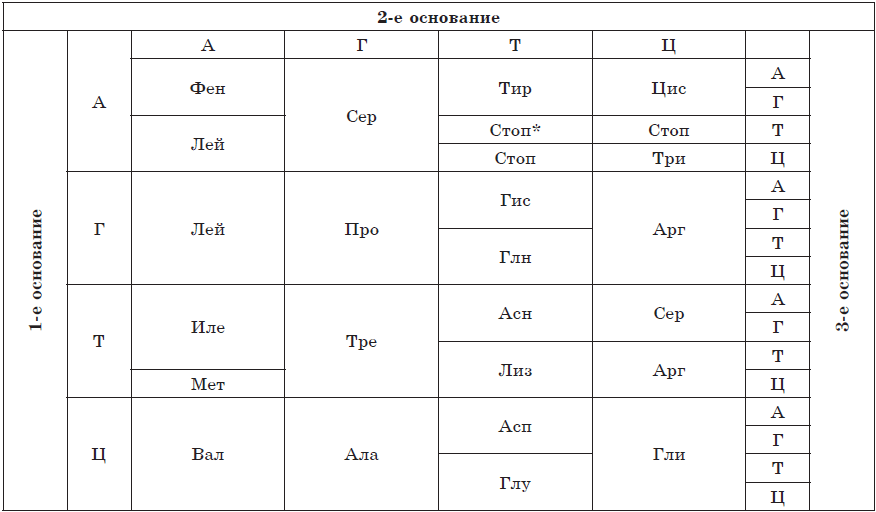
*Stop codon, indicating the end of the synthesis of the polypeptide chain.
Abbreviations for amino acid names:
Ala - alanine
Arg - arginine
Asn - asparagine
Asp - aspartic acid
Val - valine
His - histidine
Gly - glycine
Gln - glutamine
Glu - glutamic acid
Ile - isoleucine
Leu - leucine
Liz - lysine
Meth - methionine
Pro - proline
Ser - serine
Tyr - tyrosine
Tre - threonine
Three - tryptophan
Fen - phenylalanine
cis - cysteine
If you start reading genetic information not from the first nucleotide in the triplet, but from the second, then not only will the reading frame shift - the protein synthesized in this way will be completely different not only in the nucleotide sequence, but also in structure and properties. There are no punctuation marks between the triplets, so there are no obstacles to the shift of the reading frame, which opens up scope for the occurrence and maintenance of mutations.
Matrix nature of biosynthetic reactions
Bacterial cells are able to duplicate every 20-30 minutes, and eukaryotic cells - every day and even more often, which requires high speed and accuracy of DNA replication. In addition, each cell contains hundreds and thousands of copies of many proteins, especially enzymes, therefore, for their reproduction, the "piece" method of their production is unacceptable. A more progressive way is stamping, which allows you to get numerous exact copies of the product and also reduce its cost. For stamping, a matrix is needed, with which an impression is made.
In cells, the principle of matrix synthesis is that new molecules of proteins and nucleic acids are synthesized in accordance with the program laid down in the structure of pre-existing molecules of the same nucleic acids (DNA or RNA).
Biosynthesis of protein and nucleic acids
DNA replication. DNA is a double-stranded biopolymer whose monomers are nucleotides. If DNA biosynthesis proceeded according to the principle of photocopying, then numerous distortions and errors in hereditary information would inevitably arise, which would ultimately lead to the death of new organisms. Therefore, the process of DNA duplication is different, in a semi-conservative way: the DNA molecule unwinds, and on each of the chains a new chain is synthesized according to the principle of complementarity. The process of self-reproduction of the DNA molecule, which ensures the exact copying of hereditary information and its transmission from generation to generation, is called replication(from lat. replication- repetition). As a result of replication, two absolutely exact copies of the parent DNA molecule are formed, each of which carries one copy of the parent.

The process of replication is actually extremely complex, since a number of proteins are involved in it. Some of them unwind the double helix of DNA, others break hydrogen bonds between nucleotides of complementary chains, others (for example, the DNA polymerase enzyme) select new nucleotides according to the principle of complementarity, etc. Two DNA molecules formed as a result of replication diverge in two during division. newly formed daughter cells.
Errors in the replication process are extremely rare, but if they do occur, they are very quickly eliminated by both DNA polymerases and special repair enzymes, since any error in the nucleotide sequence can lead to an irreversible change in the structure and functions of the protein and, ultimately, adversely affect the viability of a new cell or even an individual.
protein biosynthesis. As the outstanding philosopher of the 19th century F. Engels figuratively put it: "Life is a form of existence of protein bodies." The structure and properties of protein molecules are determined by their primary structure, i.e., the sequence of amino acids encoded in DNA. Not only the existence of the polypeptide itself, but also the functioning of the cell as a whole depends on the accuracy of reproduction of this information; therefore, the process of protein synthesis is of great importance. It seems to be the most complex process of synthesis in the cell, since up to three hundred different enzymes and other macromolecules are involved here. In addition, it flows at a high speed, which requires even greater precision.
There are two main steps in protein biosynthesis: transcription and translation.
Transcription(from lat. transcription- rewriting) is the biosynthesis of mRNA molecules on a DNA template.

Since the DNA molecule contains two antiparallel chains, reading information from both chains would lead to the formation of completely different mRNAs, therefore their biosynthesis is possible only on one of the chains, which is called coding, or codogenic, in contrast to the second, non-coding, or non-codogenic. The rewriting process is provided by a special enzyme, RNA polymerase, which selects RNA nucleotides according to the principle of complementarity. This process can take place both in the nucleus and in organelles that have their own DNA - mitochondria and plastids.
The mRNA molecules synthesized during transcription undergo a complex process of preparation for translation (mitochondrial and plastid mRNAs can remain inside organelles, where the second stage of protein biosynthesis takes place). In the process of mRNA maturation, the first three nucleotides (AUG) and a tail of adenyl nucleotides are attached to it, the length of which determines how many protein copies can be synthesized on a given molecule. Only then do mature mRNAs leave the nucleus through nuclear pores.
In parallel, the process of amino acid activation occurs in the cytoplasm, during which the amino acid is attached to the corresponding free tRNA. This process is catalyzed by a special enzyme, it consumes ATP.
Broadcast(from lat. broadcast- transfer) is the biosynthesis of a polypeptide chain on an mRNA template, in which genetic information is translated into a sequence of amino acids of the polypeptide chain.

The second stage of protein synthesis most often occurs in the cytoplasm, for example, on the rough endoplasmic reticulum. Its occurrence requires the presence of ribosomes, activation of tRNA, during which they attach the corresponding amino acids, the presence of Mg2+ ions, as well as optimal environmental conditions (temperature, pH, pressure, etc.).
To start broadcasting initiation) a small subunit of the ribosome is attached to the mRNA molecule ready for synthesis, and then, according to the principle of complementarity, the tRNA carrying the amino acid methionine is selected to the first codon (AUG). Only then does the large subunit of the ribosome join. Within the assembled ribosome, there are two mRNA codons, the first of which is already occupied. A second tRNA, also carrying an amino acid, is attached to the codon adjacent to it, after which a peptide bond is formed between the amino acid residues with the help of enzymes. The ribosome moves one codon of the mRNA; the first of the tRNA, freed from the amino acid, returns to the cytoplasm for the next amino acid, and a fragment of the future polypeptide chain, as it were, hangs on the remaining tRNA. The next tRNA joins the new codon, which is within the ribosome, the process repeats and step by step the polypeptide chain lengthens, i.e., it elongation.
End of protein synthesis termination) occurs as soon as a specific nucleotide sequence is encountered in an mRNA molecule that does not encode an amino acid (stop codon). After that, the ribosome, mRNA and polypeptide chain are separated, and the newly synthesized protein acquires the appropriate structure and is transported to the part of the cell where it will perform its functions.
Translation is a very energy-consuming process, since the energy of one ATP molecule is spent on attaching one amino acid to tRNA, and several more are used to move the ribosome along the mRNA molecule.
To accelerate the synthesis of certain protein molecules, several ribosomes can be sequentially attached to the mRNA molecule, which form a single structure - polysome.
The cell is the genetic unit of living things. Chromosomes, their structure (shape and size) and functions. The number of chromosomes and their species constancy. Somatic and sex cells. Cell life cycle: interphase and mitosis. Mitosis is the division of somatic cells. Meiosis. Phases of mitosis and meiosis. The development of germ cells in plants and animals. Cell division is the basis for the growth, development and reproduction of organisms. The role of meiosis and mitosis
The cell is the genetic unit of life
Despite the fact that nucleic acids are the carrier of genetic information, the implementation of this information is impossible outside the cell, which is easily proved by the example of viruses. These organisms, often containing only DNA or RNA, cannot reproduce on their own, for this they must use the hereditary apparatus of the cell. They cannot even penetrate the cell without the help of the cell itself, except by using the mechanisms of membrane transport or due to cell damage. Most viruses are unstable, they die after a few hours of exposure to the open air. Therefore, the cell is a genetic unit of the living, which has a minimum set of components for the preservation, modification and implementation of hereditary information, as well as its transmission to descendants.
Most of the genetic information of a eukaryotic cell is located in the nucleus. A feature of its organization is that, unlike the DNA of a prokaryotic cell, eukaryotic DNA molecules are not closed and form complex complexes with proteins - chromosomes.
Chromosomes, their structure (shape and size) and functions
Chromosome(from Greek. chrome- color, color and catfish- body) is the structure of the cell nucleus, which contains genes and carries certain hereditary information about the signs and properties of the body.
Sometimes the ring DNA molecules of prokaryotes are also called chromosomes. Chromosomes are capable of self-duplication, they have a structural and functional individuality and retain it in a number of generations. Each cell carries all the hereditary information of the body, but only a small part of it works.
The basis of the chromosome is a double-stranded DNA molecule packed with proteins. In eukaryotes, histone and non-histone proteins interact with DNA, while in prokaryotes, histone proteins are absent.
Chromosomes are best seen under a light microscope during cell division, when, as a result of compaction, they take the form of rod-shaped bodies separated by a primary constriction - centromere — on shoulders. The chromosome may also have secondary constriction, which in some cases separates the so-called satellite. The ends of chromosomes are called telomeres. Telomeres prevent the ends of chromosomes from sticking together and ensure their attachment to the nuclear membrane in a non-dividing cell. At the beginning of division, the chromosomes are doubled and consist of two daughter chromosomes - chromatids attached at the centromere.

According to the shape, equal-arm, unequal-arm and rod-shaped chromosomes are distinguished. Chromosome sizes vary significantly, but the average chromosome has a size of 5 $×$ 1.4 µm.
In some cases, chromosomes, as a result of numerous DNA duplications, contain hundreds and thousands of chromatids: such giant chromosomes are called polythene. They are found in the salivary glands of Drosophila larvae, as well as in the digestive glands of roundworms.
The number of chromosomes and their species constancy. Somatic and germ cells
According to the cellular theory, a cell is a unit of structure, life and development of an organism. Thus, such important functions of living things as growth, reproduction and development of the organism are provided at the cellular level. Cells of multicellular organisms can be divided into somatic and sex.
somatic cells are all the cells of the body that are formed as a result of mitotic division.
The study of chromosomes made it possible to establish that the somatic cells of the organism of each biological species are characterized by a constant number of chromosomes. For example, a person has 46 of them. The set of chromosomes of somatic cells is called diploid(2n), or double.

sex cells, or gametes, are specialized cells that serve for sexual reproduction.
Gametes always contain half as many chromosomes as in somatic cells (in humans - 23), so the set of chromosomes of germ cells is called haploid(n), or single. Its formation is associated with meiotic cell division.
The amount of DNA of somatic cells is designated as 2c, and that of germ cells is 1c. The genetic formula of somatic cells is written as 2n2c, and sex - 1n1c.
In the nuclei of some somatic cells, the number of chromosomes may differ from their number in somatic cells. If this difference is greater by one, two, three, etc. haploid sets, then such cells are called polyploid(tri-, tetra-, pentaploid, respectively). In such cells, metabolic processes are usually very intensive.
The number of chromosomes in itself is not a species-specific trait, since different organisms may have the same number of chromosomes, while related ones may have different numbers. For example, malarial plasmodium and horse roundworm have two chromosomes, while humans and chimpanzees have 46 and 48, respectively.
Human chromosomes are divided into two groups: autosomes and sex chromosomes (heterochromosomes). Autosome there are 22 pairs in human somatic cells, they are the same for men and women, and sex chromosomes only one pair, but it is she who determines the sex of the individual. There are two types of sex chromosomes - X and Y. The cells of the body of a woman carry two X chromosomes, and men - X and Y.
Karyotype- this is a set of signs of the chromosome set of an organism (the number of chromosomes, their shape and size).
The conditional record of the karyotype includes the total number of chromosomes, sex chromosomes, and possible deviations in the set of chromosomes. For example, the karyotype of a normal male is written as 46,XY, while the karyotype of a normal woman is 46,XX.
Cell life cycle: interphase and mitosis
Cells do not arise each time anew, they are formed only as a result of the division of mother cells. After separation, the daughter cells take some time to form organelles and acquire the appropriate structure, which would ensure the performance of a certain function. This period of time is called ripening.
The period of time from the appearance of a cell as a result of division to its division or death is called cell life cycle.
In eukaryotic cells, the life cycle is divided into two main stages: interphase and mitosis.

Interphase- this is the period of time in the life cycle in which the cell does not divide and functions normally. The interphase is divided into three periods: G 1 -, S- and G 2 -periods.
G 1 -period(presynthetic, postmitotic) is a period of cell growth and development, during which there is an active synthesis of RNA, proteins and other substances necessary for the complete life support of the newly formed cell. By the end of this period, the cell may begin to prepare for DNA duplication.
AT S-period(synthetic) the process of DNA replication takes place. The only part of the chromosome that does not undergo replication is the centromere, therefore the resulting DNA molecules do not completely diverge, but remain fastened in it, and at the beginning of division, the chromosome has an X-shaped appearance. The genetic formula of the cell after DNA duplication is 2n4c. Also in the S-period, doubling of the centrioles of the cell center occurs.
G 2 -period(postsynthetic, premitotic) is characterized by intensive synthesis of RNA, proteins and ATP necessary for the process of cell division, as well as the separation of centrioles, mitochondria and plastids. Until the end of the interphase, the chromatin and nucleolus remain clearly distinguishable, the integrity of the nuclear membrane is not disturbed, and the organelles do not change.
Some of the cells of the body are able to perform their functions throughout the life of the body (neurons of our brain, muscle cells of the heart), while others exist for a short time, after which they die (cells of the intestinal epithelium, cells of the epidermis of the skin). Consequently, the processes of cell division and the formation of new cells must constantly occur in the body, which would replace the dead ones. Cells capable of dividing are called stem. In the human body, they are found in the red bone marrow, in the deep layers of the epidermis of the skin and other places. Using these cells, you can grow a new organ, achieve rejuvenation, and also clone the body. The prospects for the use of stem cells are quite clear, but the moral and ethical aspects of this problem are still being discussed, since in most cases embryonic stem cells obtained from human fetuses killed during abortion are used.
The duration of interphase in plant and animal cells averages 10-20 hours, while mitosis takes about 1-2 hours.
In the course of successive divisions in multicellular organisms, daughter cells become more and more diverse, since they read information from an increasing number of genes.
Some cells eventually stop dividing and die, which may be due to the completion of certain functions, as in the case of epidermal cells of the skin and blood cells, or to damage to these cells by environmental factors, in particular pathogens. Genetically programmed cell death is called apoptosis while accidental death is necrosis.
Mitosis is the division of somatic cells. Phases of mitosis
Mitosis- a method of indirect division of somatic cells.
During mitosis, the cell goes through a series of successive phases, as a result of which each daughter cell receives the same set of chromosomes as in the mother cell.
Mitosis is divided into four main phases: prophase, metaphase, anaphase, and telophase. Prophase- the longest stage of mitosis, during which chromatin condensation occurs, as a result of which X-shaped chromosomes, consisting of two chromatids (daughter chromosomes), become visible. In this case, the nucleolus disappears, the centrioles diverge towards the poles of the cell, and the achromatin spindle (spindle) of microtubules begins to form. At the end of prophase, the nuclear membrane breaks up into separate vesicles.

AT metaphase chromosomes line up along the equator of the cell with their centromeres, to which microtubules of a fully formed division spindle are attached. At this stage of division, the chromosomes are most dense and have a characteristic shape, which makes it possible to study the karyotype.
AT anaphase rapid DNA replication occurs in the centromeres, as a result of which the chromosomes split and the chromatids diverge towards the poles of the cell, stretched by microtubules. The distribution of chromatids must be absolutely equal, since it is this process that maintains the constancy of the number of chromosomes in the cells of the body.
On the stage telophase daughter chromosomes gather at the poles, despiralize, around them nuclear envelopes form from the vesicles, and nucleoli appear in the newly formed nuclei.
After the division of the nucleus, the division of the cytoplasm occurs - cytokinesis, during which there is a more or less uniform distribution of all the organelles of the mother cell.
Thus, as a result of mitosis, two daughter cells are formed from one mother cell, each of which is a genetic copy of the mother cell (2n2c).
In diseased, damaged, aging cells and specialized tissues of the body, a slightly different division process can occur - amitosis. Amitosis called the direct division of eukaryotic cells, in which the formation of genetically equivalent cells does not occur, since the cellular components are distributed unevenly. It occurs in plants in the endosperm and in animals in the liver, cartilage, and cornea of the eye.
Meiosis. Phases of meiosis
Meiosis- this is a method of indirect division of primary germ cells (2n2c), as a result of which haploid cells (1n1c), most often germ cells, are formed.
Unlike mitosis, meiosis consists of two successive cell divisions, each preceded by an interphase. The first division of meiosis (meiosis I) is called reduction, since in this case the number of chromosomes is halved, and the second division (meiosis II) - equational, since in its process the number of chromosomes is conserved.

Interphase I proceeds similarly to the interphase of mitosis. Meiosis I is divided into four phases: prophase I, metaphase I, anaphase I and telophase I. prophase I Two major processes occur: conjugation and crossing over. Conjugation- this is the process of fusion of homologous (paired) chromosomes along the entire length. The pairs of chromosomes formed during conjugation are retained until the end of metaphase I.
Crossing over- mutual exchange of homologous regions of homologous chromosomes. As a result of crossing over, the chromosomes received by the organism from both parents acquire new combinations of genes, which leads to the appearance of genetically diverse offspring. At the end of prophase I, as in the prophase of mitosis, the nucleolus disappears, the centrioles diverge towards the poles of the cell, and the nuclear envelope disintegrates.

AT metaphase I pairs of chromosomes line up along the equator of the cell, microtubules of the fission spindle are attached to their centromeres.
AT anaphase I whole homologous chromosomes consisting of two chromatids diverge to the poles.
AT telophase I around clusters of chromosomes at the poles of the cell, nuclear membranes form, nucleoli form.
Cytokinesis I provides division of cytoplasms of daughter cells.
The daughter cells formed as a result of meiosis I (1n2c) are genetically heterogeneous, since their chromosomes, randomly dispersed to the poles of the cell, contain unequal genes.
Comparative characteristics of mitosis and meiosis
| sign | Mitosis | Meiosis | |
| What cells start dividing? | Somatic (2n) | Primary germ cells (2n) | |
| Number of divisions | 1 | 2 | |
| How many and what kind of cells are formed in the process of division? | 2 somatic (2n) | 4 sexual (n) | |
| Interphase | Cell preparation for division, DNA duplication | Very short, DNA duplication does not occur | |
| Phases | Meiosis I | Meiosis II | |
| Prophase | Chromosome condensation, disappearance of the nucleolus, disintegration of the nuclear envelope, conjugation and crossing over may occur | Condensation of chromosomes, disappearance of the nucleolus, disintegration of the nuclear envelope | |
| metaphase | Pairs of chromosomes are located along the equator, a division spindle is formed | Chromosomes line up along the equator, the spindle of division is formed | |
| Anaphase | Homologous chromosomes from two chromatids diverge towards the poles | Chromatids diverge towards the poles | |
| Telophase | Chromosomes despiralize, new nuclear envelopes and nucleoli form | Chromosomes despiralize, new nuclear envelopes and nucleoli form | |
Interphase II very short, since DNA doubling does not occur in it, that is, there is no S-period.
Meiosis II also divided into four phases: prophase II, metaphase II, anaphase II and telophase II. AT prophase II the same processes occur as in prophase I, with the exception of conjugation and crossing over.
AT metaphase II Chromosomes are located along the equator of the cell.
AT anaphase II Chromosomes split at the centromere and the chromatids stretch towards the poles.
AT telophase II nuclear membranes and nucleoli form around clusters of daughter chromosomes.
After cytokinesis II the genetic formula of all four daughter cells is 1n1c, but they all have a different set of genes, which is the result of crossing over and a random combination of maternal and paternal chromosomes in daughter cells.
The development of germ cells in plants and animals
Gametogenesis(from Greek. gamete- wife, gametes- husband and genesis- origin, occurrence) is the process of formation of mature germ cells.
Since sexual reproduction most often requires two individuals - female and male, producing different sex cells - eggs and sperm, then the processes of formation of these gametes should be different.
The nature of the process also depends to a large extent on whether it occurs in a plant or animal cell, since in plants only mitosis occurs during the formation of gametes, while in animals both mitosis and meiosis occur.
The development of germ cells in plants. In angiosperms, the formation of male and female germ cells occurs in different parts of the flower - stamens and pistils, respectively.
Before the formation of male germ cells - microgametogenesis(from Greek. micros- small) - happening microsporogenesis, that is, the formation of microspores in the anthers of the stamens. This process is associated with the meiotic division of the mother cell, which results in four haploid microspores. Microgametogenesis is associated with the mitotic division of microspores, giving a male gametophyte of two cells - a large vegetative(siphonogenic) and shallow generative. After division, the male gametophyte is covered with dense shells and forms a pollen grain. In some cases, even in the process of pollen maturation, and sometimes only after transfer to the stigma of the pistil, the generative cell divides mitotically with the formation of two immobile male germ cells - sperm. After pollination, a pollen tube is formed from the vegetative cell, through which sperm penetrate into the ovary of the pistil for fertilization.

The development of female germ cells in plants is called megagametogenesis(from Greek. megas- big). It occurs in the ovary of the pistil, which is preceded by megasporogenesis, as a result of which four megaspores are formed from the mother cell of the megaspore lying in the nucellus by meiotic division. One of the megaspores divides mitotically three times, giving rise to the female gametophyte, an embryo sac with eight nuclei. With the subsequent isolation of the cytoplasms of the daughter cells, one of the resulting cells becomes an egg, on the sides of which lie the so-called synergids, three antipodes are formed at the opposite end of the embryo sac, and in the center, as a result of the fusion of two haploid nuclei, a diploid central cell is formed.

The development of germ cells in animals. In animals, two processes of the formation of germ cells are distinguished - spermatogenesis and oogenesis.
spermatogenesis(from Greek. sperm, spermatos- seed and genesis- origin, occurrence) is the process of formation of mature male germ cells - spermatozoa. In humans, it occurs in the testes, or testes, and is divided into four periods: reproduction, growth, maturation and formation.

AT breeding season primordial germ cells divide mitotically, resulting in the formation of diploid spermatogonia. AT growth period spermatogonia accumulate nutrients in the cytoplasm, increase in size and turn into primary spermatocytes, or spermatocytes of the 1st order. Only after that they enter meiosis ( ripening period), which first results in two secondary spermatocyte, or spermatocyte of the 2nd order, and then - four haploid cells with a fairly large amount of cytoplasm - spermatids. AT formation period they lose almost all of the cytoplasm and form a flagellum, turning into spermatozoa.
spermatozoa, or gummies, - very small mobile male sex cells with a head, neck and tail.

AT head, except for the core, is acrosome- a modified Golgi complex, which ensures the dissolution of the membranes of the egg during fertilization. AT neck there are centrioles of the cell center, and the basis ponytail form microtubules that directly support the movement of the spermatozoon. It also contains mitochondria, which provide the sperm with ATP energy for movement.
Ovogenesis(from Greek. UN- an egg and genesis- origin, occurrence) is the process of formation of mature female germ cells - eggs. In humans, it occurs in the ovaries and consists of three periods: reproduction, growth and maturation. Periods of reproduction and growth, similar to those in spermatogenesis, occur even during intrauterine development. At the same time, diploid cells are formed from the primary germ cells as a result of mitosis. oogonia, which then turn into diploid primary oocytes, or oocytes of the 1st order. Meiosis and subsequent cytokinesis occurring in ripening period, are characterized by uneven division of the cytoplasm of the mother cell, so that as a result, at first one is obtained secondary oocyte, or oocyte 2nd order, and first polar body, and then from the secondary oocyte - the egg, which retains the entire supply of nutrients, and the second polar body, while the first polar body is divided into two. Polar bodies take away excess genetic material.
In humans, eggs are produced with an interval of 28-29 days. The cycle associated with the maturation and release of eggs is called the menstrual cycle.
Egg- a large female germ cell, which carries not only a haploid set of chromosomes, but also a significant supply of nutrients for the subsequent development of the embryo.

The egg in mammals is covered with four membranes, which reduce the likelihood of damage to it by various factors. The diameter of the egg in humans reaches 150-200 microns, while in an ostrich it can be several centimeters.
Cell division is the basis for the growth, development and reproduction of organisms. The role of mitosis and meiosis
If in unicellular organisms cell division leads to an increase in the number of individuals, i.e., reproduction, then in multicellular organisms this process can have a different meaning. Thus, cell division of the embryo, starting from the zygote, is the biological basis for the interconnected processes of growth and development. Similar changes are observed in a person during adolescence, when the number of cells not only increases, but also a qualitative change in the body occurs. Reproduction of multicellular organisms is also based on cell division, for example, during asexual reproduction, due to this process, a whole body is restored from a part of the body, and during sexual reproduction, germ cells are formed during gametogenesis, subsequently giving a new organism. It should be noted that the main methods of eukaryotic cell division—mitosis and meiosis—have different significance in the life cycles of organisms.
As a result of mitosis, there is a uniform distribution of hereditary material between daughter cells - exact copies of the mother. Without mitosis, the existence and growth of multicellular organisms developing from a single cell, the zygote, would be impossible, since all cells of such organisms must contain the same genetic information.
In the process of division, daughter cells become more and more diverse in structure and functions, which is associated with the activation of new groups of genes in them due to intercellular interaction. Thus, mitosis is necessary for the development of an organism.
This method of cell division is necessary for the processes of asexual reproduction and regeneration (recovery) of damaged tissues, as well as organs.
Meiosis, in turn, ensures the constancy of the karyotype during sexual reproduction, as it reduces by half the set of chromosomes before sexual reproduction, which is then restored as a result of fertilization. In addition, meiosis leads to the appearance of new combinations of parental genes due to crossing over and random combination of chromosomes in daughter cells. Thanks to this, the offspring is genetically diverse, which provides material for natural selection and is the material basis of evolution. A change in the number, shape and size of chromosomes, on the one hand, can lead to the appearance of various deviations in the development of the organism and even its death, and on the other hand, it can lead to the appearance of individuals more adapted to the environment.
Thus, the cell is a unit of growth, development and reproduction of organisms.
Cytology as a science.
The history of cytology is closely connected with the invention, use and improvement of the microscope.
1665: R. Hooke, observing for the first time under a microscope a thin section of a cork tree, discovered empty cells, which he called celluli , or cells; in fact, R. Hooke observed only the shells of plant cells.
1671: N. Grew, M. Malpighi, studying the anatomy of plants, also discovered the smallest "cells", "vesicles" or "sacs".
1674: A. Van Leeuwenhoek first discovered, and then repeatedly observed under a microscope in a drop of water, unicellular animal organisms.
During this period, its wall was considered the main part of the cell, and only two hundred years later it became clear that the main thing in the cell is not the wall, but the internal contents. In the 18th century, new information about the cell accumulated slowly, and in the field of zoology more slowly than in botany, since real cell walls, which served as the main subject of study, are characteristic only of plant cells. In relation to animal cells, scientists did not dare to apply this term and identify them with plant cells.
In the future, as the microscope and microscopy techniques were improved, information about animal and plant cells was also accumulated. Already by the 30s of the 19th century, a lot of information on cell morphology had accumulated, and it was found that the cytoplasm and nucleus are its essential components:
1802, 1808: C. Brissot-Mirbe established the fact that all plant organisms are formed by tissues, which consist of cells.
1809: J. B. Lamarck extended Brissot-Mirbe's idea of cellular structure to animals.
1825: J. Purkinė discovered protoplasm, a semi-liquid gelatinous content of cells.
1831: R. Brown discovered the nucleus in plant cells.
1833: R. Brown came to the conclusion that the nucleus is an essential part of the plant cell.
1839: T. Schwann summarized all the data accumulated by that time and formulated the cellular theory.
1855: R. Virchow proved that all cells are formed from other cells by division.
1866: Haeckel established that the preservation and transmission of hereditary traits is carried out by the nucleus.
1866-1898: Describes the main components of a cell that can be seen under an optical microscope. Cytology acquires the character of an experimental science.
1900: Following the advent of genetics, cytogenetics begins to develop, studying the behavior of chromosomes during division and fertilization.
1946: The use of the electron microscope began in biology, which made it possible to study the ultrastructures of cells.
Cytology - a science that studies the structure, chemical composition and functions of cells, their reproduction, development and interaction in a multicellular organism.
The subject of cytology- cells of unicellular and multicellular prokaryotic and eukaryotic organisms.
Tasks of cytology:
1. Study of the structure and functions of cells and their components (membranes, organelles, inclusions, nuclei).
2. The study of the chemical composition of cells, biochemical reactions occurring in them.
3. The study of the relationship of cells of a multicellular organism.
4. Study of cell division.
5. Studying the possibility of cell adaptation to environmental changes.
To solve the tasks in cytology, various methods are used.
Microscopic methods: allow you to study the structure of the cell and its components using microscopes (light, phase-contrast, luminescent, ultraviolet, electron); light microscopy is based on the flow of light; studies cells and their large structures; electron microscopy - the study of small structures (membranes, ribosomes, etc.) in an electron beam with a wavelength less than that of visible light.
Cyto- and histochemical methods- based on the selective action of reagents and dyes on certain substances of the cytoplasm; used to establish the chemical composition and localization of various components (proteins, DNA, RNA, lipids, etc.) in cells.
Histological method is a method of preparation of micropreparations from native and fixed tissues and organs. The native material is frozen, and the fixed object goes through the stages of compaction, pouring into paraffin. The test material is then sectioned, stained and placed in Canadian balsam.
Biochemical methods allow you to study the chemical composition of cells and the biochemical reactions occurring in them.
Differential centrifugation method: based on different sedimentation rates of cell components; selects individual cell components (mitochondria, ribosomes, etc.) for further study by other methods.
Method of X-ray diffraction analysis: after the introduction of metal atoms into the cell, the spatial configuration and some physical properties of macromolecules (protein, DNA) are studied.
Autoradiography method– introduction of radioactive (labeled) isotopes into the cell and further study of their incorporation into substances synthesized by the cell; allows you to study the processes of matrix synthesis and cell division.
Film and photography method fix the processes of cell division.
Microsurgical methods allow the transplantation of cell components (organelles, nucleus) from one cell to another in order to study their functions.
Cell culture method– cultivation of individual cells on nutrient media under sterile conditions; makes it possible to study the division, differentiation and specialization of cells, to obtain clones of plant organisms.
Knowledge of the basics of chemical and structural organization, principles of functioning and mechanisms of cell development is extremely important for understanding the similarities inherent in the complex organisms of plants, animals and humans. The development of the IVF method is an example of the practical application of cytological knowledge.
Cell theory. The main provisions of modern cell theory.
cell theory - a scientific generalization in biology, according to which the cell is recognized as a common structural unit of living organisms, the similarity of animal and plant cells in structure, function and development is affirmed. The cellular theory reduces the structure of the most complex living beings to the structure of cells, their development - to the reproduction, growth and development of cells.
“It was only from the time of this discovery that the study of organic, living products of nature, both comparative anatomy and physiology, and embryology, became on solid ground. The veil of mystery that shrouded the process of emergence and growth and the structure of organisms was torn off. An incomprehensible miracle appeared in the form of a process occurring according to the law identical for all multicellular organisms ”(F. Engels). Independently of each other, the essence of the cellular theory was stated in their works by M. Schleiden "Data on the development of plants" (1838) and T. Schwann "Microscopic studies on the correspondence in the structure and growth of animals and plants" (1839):
1. The cell is the main structural unit of all plant and animal organisms.
2. The process of cell formation determines the growth (development and differentiation) of plant and animal tissues.
3. A cell within certain limits is an individual, a kind of independent whole, and an organism is a kind of their sum.
4. New cells arise from cytoblastoma.
The first two conclusions remain relevant today.
Although the creation of the cell theory is associated with the names of Schleiden and Schwann, the idea of the unity of the structure of plants and animals was repeatedly expressed by Lamarck (1809), Dutrochet (1824), Mol (1831), Goryaninov.
In 1858, Rudolf Virchow in his work "Cellular Pathology":
1. Showed the connection of pathological processes with morphological structures, with certain changes in the structure of cells; the disease of the whole organism is determined by the disease of the cell.
2. Instead of T. Schwann's thesis about cytoblastoma, another one puts forward: Omnis cellula ex cellule - each cell is from a cell.
3. He suggested that there is no life outside of cells.
Virchow R. also considered the body as the sum of its constituent cells, which was criticized by I. M. Sechenov, S. P. Botkin and I. P. Pavlov. They showed that a multicellular organism is a single whole and the activity of organisms, as well as the integration of its parts, is carried out primarily by the central nervous system. In the XIX - XX centuries. thanks to the use of more modern methods of cytological analysis, new data were obtained (the complex structure of the cell, its main organelles, methods of cell division, etc. were described), which made it possible to confirm, clarify and supplement the cell theory:
All living organisms are made up of cells (with the exception of viruses);
Cells of unicellular and multicellular organisms are similar (homologous) in structure, chemical composition, principles of metabolism and basic manifestations of vital activity;
It is the cell that has the whole set of features that characterize the living;
All living organisms develop from one or from a group of cells;
Each cell is formed as a result of the division of the original (mother) cell;
In complex multicellular organisms, cells differentiate by specializing in a particular function;
Cells are combined into tissues and organs, functionally connected systems and are under the control of intercellular, humoral and nervous forms of regulation.
The main provisions of modern cell theory:
1. A cell is an elementary unit of a living thing, capable of self-renewal, self-regulation and self-reproduction; is a unit of structure, functioning and development of all living organisms.
2. The cells of all living organisms are similar in structure, chemical composition and basic manifestations of vital activity.
3. Cells are formed by dividing the original (mother) cell.
4. In a multicellular organism, cells specialize in functions and form tissues from which organs and organ systems are built, interconnected by intercellular, humoral and nervous forms of regulation.
Thus, the creation of the cellular theory became the most important event in natural science, one of the decisive proofs of the unity of living nature. The cell theory had a significant impact on the development of biology and served as the foundation for the further development of many biological disciplines - embryology, histology, physiology, etc.
Cell structure.
If the cells of bacteria and other prokaryotes are relatively simple in structure and carry a number of primitive features inherited from the first living organisms on Earth, then eukaryotic cells - from protozoa (protists) to cells of higher plants and mammals - differ in complexity and diversity of structure. There is no such thing as a typical cell, but there are some commonalities among the thousands of different types of cells.
Cells of tissues of plants, fungi and animals, depending on the functions they perform, have not only different sizes, but also different shapes. The diameter of most eukaryotic cells is 10-100 microns, the smallest cells are about 4 microns in size, some have 1-10 mm (watermelon pulp cells), and the largest (ostrich, penguin, geese eggs) 10-20 cm, sometimes more ( processes of nerve cells can reach 1 meter). By shape, cells can be distinguished: round, polygonal, rod-shaped, stellate (nerve), disc-shaped (erythrocytes), cylindrical, cubic, etc.
The cell consists of three structural components - the shell (plasmalemma), cytoplasm and nucleus (Fig ....).
CELL STRUCTURE
Shell Cytoplasm Nucleus
lipid bilayer; - hyaloplasm; - nuclear membrane;
Two layers of proteins; - common organelles - nuclear juice;
appointments;
Oligosaccharides; - special organelles - chromatin;
appointments; - nucleolus.
Inclusions.
Fig.1. A generalized scheme of the structure of a eukaryotic cell.
4. Biological membranes. Cytoplasmic membrane: structure, properties, functions.
Cells are characterized by a membrane principle of structure.
biological membrane - thin films, protein-lipid structure, 7 - 10 nm thick, located on the surface of cells (cell membrane), forming the walls of most organelles and the shell of the nucleus.
In 1972, S. Singer and G. Nichols proposed fluid mosaic model structure of the cell membrane. Later it was practically confirmed. When viewed under an electron microscope, three layers can be seen. Medium, light, forms the basis of the membrane - the bilipid layer formed by liquid phospholipids ("lipid sea"). Molecules of membrane lipids (phospholipids, glycolipids, cholesterol, etc.) have hydrophilic heads and hydrophobic tails, therefore they are orderly oriented in the bilayer. The two dark layers are proteins that are located differently relative to the lipid bilayer: peripheral (adjacent) - most proteins are located on both surfaces of the lipid layer; semi-integral (semi-submerged) - penetrate only one layer of lipids; integral (submerged) pass through both layers. Proteins have hydrophobic regions that interact with lipids, and hydrophilic regions on the surface of the membrane in contact with the aqueous contents of the cell, or tissue fluid.
Functions of biological membranes:
1) delimits the contents of the cell from the external environment and the contents of organelles, the nucleus from
2) provide transport of substances into and out of the cell into the cytoplasm from organelles and vice versa;
3) participate in receiving and converting signals from the environment, recognizing cell substances, etc.;
4) provide near-membrane processes;
5) participate in the transformation of energy.
Cytoplasmic membrane (plasmalemma, cell membrane, plasma membrane) - the biological membrane that surrounds the cell. Its thickness is about 7.5 nm. It has a structure characteristic of biological membranes. On the surface of the membrane there are oligosaccharide chains (antennas) that perform the following functions: recognition of external signals; adhesion of cells, their correct orientation during tissue formation; immune response, where glycoproteins play the role of an immune response.
The chemical composition of the plasma membrane: 55% - proteins, 35% - lipids, 2-10% - oligosaccharides.
The outer cell membrane forms a mobile surface of the cell, which can have outgrowths and protrusions, performs undulating oscillatory movements, macromolecules are constantly moving in it. The cell surface is heterogeneous: its structure is not the same in different areas, and the physiological properties of these areas are also not the same. Some enzymes (about 200) are localized in the plasmalemma, so the effect of environmental factors on the cell is mediated by its cytoplasmic membrane. The surface of the cell has high strength and elasticity, it is easily and quickly restored after minor damage.
The structure of the plasma membrane determines its properties:
Plasticity (fluidity), allows the membrane to change its shape and size;
The ability to self-closing, enables the membrane to restore integrity when ruptured;
Selective permeability provides the passage of various substances through the membrane at different speeds.
The main functions of the cytoplasmic membrane:
determines and maintains the shape of the cell;
Delimits the internal contents of the cell;
protects the cell from mechanical influences and penetration of damaging biological agents;
Regulates the metabolism between the cell and the environment, ensuring the constancy of the intracellular composition;
recognizes external signals, “recognizes” certain substances (for example, hormones);
participates in the formation of intercellular contacts and various kinds of specific protrusions of the cytoplasm (microvilli, cilia, flagella).
Discovery and study cells made possible by the invention of the microscope and the improvement of microscopic examination methods.
In 1665, the Englishman Robert Hooke was the first to observe the division of cork oak bark tissue into cells (cells) using magnifying lenses. Although it turned out that he did not discover cells (in his own concept of the term), but only the outer shells of plant cells. Later, the world of unicellular organisms was discovered by A. Leeuwenhoek. He was the first to see animal cells (erythrocytes). Later, F. Fontana described animal cells, but these studies at that time did not lead to the concept of the universality of the cellular structure, because there were no clear ideas about what a cell is.
R. Hooke believed that cells are voids or pores between plant fibers. Later, M. Malpighi, N. Gru and F. Fontana, observing plant objects under a microscope, confirmed the data of R. Hooke, calling the cells “bubbles”. A. Levenguk made a significant contribution to the development of microscopic studies of plant and animal organisms. He published the data of his observations in the book "Secrets of Nature".
The illustrations for this book clearly demonstrate the cellular structures of plant and animal organisms. However, A. Leeuwenhoek did not represent the described morphological structures as cellular formations. His research was random, not systematized. G. Link, G. Travenarius and K. Rudolph at the beginning of the $19th century showed by their research that cells are not voids, but independent formations limited by walls. It was found that the cells have contents that I called protoplasm Purkinje. R. Brown described the nucleus as a permanent part of the cells.
T. Schwann analyzed the literature data on the cellular structure of plants and animals, comparing them with his own research and published the results in his work. In it, T. Schwann showed that cells are elementary living structural units of plant and animal organisms. They have a common structural plan and are formed in a single way. These theses became the basis of the cell theory.
Researchers for a long time were engaged in the accumulation of observations of the structure of unicellular and multicellular organisms, before formulating the provisions of CT. It was during this period that various optical research methods were more developed and improved.
Cells are divided into nuclear (eukaryotic) and non-nuclear (prokaryotic). Animals are built from eukaryotic cells. Only mammalian red blood cells (erythrocytes) do not have nuclei. They lose them in the course of their development.
The definition of a cell has changed depending on the knowledge of their structure and function.
Definition 1
According to modern data, cell - this is a structurally ordered system of biopolymers limited by the active shell, which form the nucleus and cytoplasm, participate in a single set of metabolic processes and ensure the maintenance and reproduction of the system as a whole.
cell theory is a generalized idea of the structure of the cell as a unit of life, of the reproduction of cells and their role in the formation of multicellular organisms.
Progress in the study of cells is associated with the development of microscopy in the $19th century. At that time, the idea of the structure of the cell changed: not the cell membrane was taken as the basis of the cell, but its contents - protoplasm. At the same time, the nucleus was discovered as a permanent element of the cell.
Information about the fine structure and development of tissues and cells made it possible to generalize. Such a generalization was made in 1839 by the German biologist T. Schwann in the form of the cell theory formulated by him. He argued that the cells of both animals and plants are fundamentally similar. The German pathologist R. Virchow developed and generalized these ideas. He put forward an important position, which was that cells arise only from cells by reproduction.
Basic provisions of cell theory
T. Schwann in 1839, in his work “Microscopic studies on the correspondence in the structure and growth of animals and plants”, he formulated the main provisions of the cell theory (later they were refined and supplemented more than once.
The cell theory contains the following provisions:
- cell - the basic elementary unit of the structure, development and functioning of all living organisms, the smallest unit of life;
- cells of all organisms are homologous (similar) (homologous) in their chemical structure, the main manifestations of life processes and metabolism;
- cells multiply by division - a new cell is formed as a result of the division of the original (mother) cell;
- in complex multicellular organisms, cells specialize in the functions they perform and form tissues; organs are built from tissues, closely interconnected by intercellular, humoral and nervous forms of regulation.
The intensive development of cytology in the $XIX$ and $XX$ centuries confirmed the main provisions of CT and enriched it with new data on the structure and functions of the cell. During this period, some incorrect theses of the cellular theory of T. Schwann were discarded, namely, that a single cell of a multicellular organism can function independently, that a multicellular organism is a simple collection of cells, and the development of a cell occurs from a non-cellular “blastema”.
In its modern form, cell theory includes the following main provisions:
- A cell is the smallest unit of a living thing, which has all the properties that meet the definition of "living". These are metabolism and energy, movement, growth, irritability, adaptation, variability, reproduction, aging and death.
- The cells of various organisms have a common structural plan, which is due to the similarity of the general functions aimed at maintaining the life of the cells themselves and their reproduction. The diversity of cell forms is the result of the specificity of their functions.
- Cells multiply as a result of the division of the original cell with the previous reproduction of its genetic material.
- Cells are parts of an integral organism, their development, structural features and functions depend on the whole organism, which is a consequence of the interaction in the functional systems of tissues, organs, apparatuses and organ systems.
Remark 1
The cell theory, which corresponds to the current level of knowledge in biology, in many respects radically differs from the ideas about the cell not only at the beginning of the 19th century, when T. Schwann formulated it for the first time, but even in the middle of the 20th century. In our time, this is a system of scientific views, which has taken the form of theories, laws and principles.
The main provisions of CT have retained their significance to this day, although for more than 150 years new information has been obtained on the structure, vital activity and development of cells.
Significance of cell theory
The significance of the cell theory in the development of science lies in the fact that thanks to it it became clear that the cell is the most important component of all organisms, their main "building" component. Since the development of each organism begins with a single cell (zygote), the cell is also the embryonic basis of multicellular organisms.
The creation of the cell theory has become one of the decisive proofs of the unity of all living nature, the most important event in biological science.
Cell theory contributed to the development of embryology, histology and physiology. It provided the basis for the materialistic concept of life, for explaining the evolutionary interconnection of organisms, for the concept of the essence of ontogeny.
The main provisions of CT are still relevant today, although over a period of more than 100 years, natural scientists have received new information about the structure, development and life of the cell.
The cell is the basis of all processes in the body: both biochemical and physiological, since it is at the cellular level that all these processes occur. Thanks to the cellular theory, it became possible to come to the conclusion about the similarity in the chemical composition of all cells and once again to be convinced of the unity of the entire organic world.
The cell theory is one of the most important biological generalizations, according to which all organisms have a cellular structure.
Remark 2
The cellular theory, together with the law of energy transformation and the evolutionary theory of Charles Darwin, is one of the three greatest discoveries of natural science in the $19th century.
Cell theory has dramatically influenced the development of biology. She proved the unity of living nature and showed the structural unit of this unity, which is the cell.
The creation of the cell theory has become a major event in biology, one of the decisive proofs of the unity of all living nature. The cell theory had a significant and decisive influence on the development of biology, serving as the main foundation for the development of such disciplines as embryology, histology and physiology. It provided a basis for explaining the related relationships of organisms, for the concept of the mechanism of individual development.
Cell theory is perhaps the most important generalization of modern biology and is a system of principles and provisions. It is the scientific background for many biological disciplines that study the structure and life of living beings. The cell theory reveals the mechanisms of growth, development and reproduction of organisms.
) supplemented it with the most important provision (every cell comes from another cell).
Schleiden and Schwann, summarizing the available knowledge about the cell, proved that the cell is the basic unit of any organism. Animal cells, plants and bacteria have a similar structure. Later, these conclusions became the basis for proving the unity of organisms. T. Schwann and M. Schleiden introduced the fundamental concept of the cell into science: there is no life outside the cells. The cellular theory was supplemented and edited every time.
Provisions of the cell theory of Schleiden-Schwann
- All animals and plants are made up of cells.
- Plants and animals grow and develop through the formation of new cells.
- A cell is the smallest unit of life, and the whole organism is a collection of cells.
The main provisions of modern cell theory
- The cell is the elementary unit of life; there is no life outside the cell.
- A cell is a single system, it includes many naturally interconnected elements, representing a holistic formation, consisting of conjugated functional units - organelles.
- The cells of all organisms are homologous.
- The cell occurs only by dividing the mother cell, after doubling its genetic material.
- A multicellular organism is a complex system of many cells united and integrated into systems of tissues and organs connected with each other.
- The cells of multicellular organisms are totipotent.
Additional Provisions of Cell Theory
In order to bring the cellular theory more fully into line with the data of modern cell biology, the list of its provisions is often supplemented and expanded. In many sources, these additional provisions differ, their set is quite arbitrary.
- Prokaryotic and eukaryotic cells are systems of different levels of complexity and are not completely homologous to each other (see below).
- The basis of cell division and reproduction of organisms is the copying of hereditary information - nucleic acid molecules ("each molecule from a molecule"). The provisions on genetic continuity apply not only to the cell as a whole, but also to some of its smaller components - to mitochondria, chloroplasts, genes and chromosomes.
- A multicellular organism is a new system, a complex ensemble of many cells united and integrated in a system of tissues and organs, connected to each other by chemical factors, humoral and nervous (molecular regulation).
- Multicellular cells are totipotent, that is, they have the genetic potencies of all cells of a given organism, are equivalent in genetic information, but differ from each other in different expression (work) of various genes, which leads to their morphological and functional diversity - to differentiation.
Story
17th century
Link and Moldenhower establish that plant cells have independent walls. It turns out that the cell is a kind of morphologically isolated structure. In 1831, Mol proves that even seemingly non-cellular plant structures, like aquifers, develop from cells.
Meyen in "Phytotomy" (1830) describes plant cells that "are either solitary, so that each cell is a separate individual, as is found in algae and fungi, or, forming more highly organized plants, they are combined into more or less significant masses. Meyen emphasizes the independence of the metabolism of each cell.
In 1831, Robert Brown describes the nucleus and suggests that it is a permanent part of the plant cell.
Purkinje School
In 1801, Vigia introduced the concept of animal tissues, but he isolated tissues on the basis of anatomical preparation and did not use a microscope. The development of ideas about the microscopic structure of animal tissues is associated primarily with the research of Purkinje, who founded his school in Breslau.
Purkinje and his students (G. Valentin should be especially noted) revealed in the first and most general form the microscopic structure of tissues and organs of mammals (including humans). Purkinje and Valentin compared individual plant cells with particular microscopic animal tissue structures, which Purkinje most often called "seeds" (for some animal structures, the term "cell" was used in his school).
In 1837 Purkinje delivered a series of lectures in Prague. In them, he reported on his observations on the structure of the gastric glands, the nervous system, etc. In the table attached to his report, clear images of some cells of animal tissues were given. Nevertheless, Purkinje could not establish the homology of plant cells and animal cells:
- firstly, by grains he understood either cells or cell nuclei;
- secondly, the term "cell" was then understood literally as "a space bounded by walls."
Purkinje compared plant cells and animal "seeds" in terms of analogy, not homology of these structures (understanding the terms "analogy" and "homology" in the modern sense).
Müller school and Schwann's work
The second school where the microscopic structure of animal tissues was studied was the laboratory of Johannes Müller in Berlin. Müller studied the microscopic structure of the dorsal string (chord); his student Henle published a study on the intestinal epithelium, in which he gave a description of its various types and their cellular structure.
Here the classic studies of Theodor Schwann were carried out, laying the foundation for the cell theory. Schwann's work was strongly influenced by the school of Purkinje and Henle. Schwann found the correct principle for comparing plant cells and the elementary microscopic structures of animals. Schwann was able to establish homology and prove correspondence in the structure and growth of the elementary microscopic structures of plants and animals.
The significance of the nucleus in the Schwann cell was prompted by the research of Matthias Schleiden, who in 1838 published the work Materials on Phytogenesis. Therefore, Schleiden is often called a co-author of the cell theory. The basic idea of the cell theory - the correspondence of plant cells and the elementary structures of animals - was alien to Schleiden. He formulated the theory of new cell formation from a structureless substance, according to which, first, the nucleolus condenses from the smallest granularity, and a nucleus is formed around it, which is the cell's former (cytoblast). However, this theory was based on incorrect facts.
In 1838, Schwann published 3 preliminary reports, and in 1839 his classic work “Microscopic studies on the correspondence in the structure and growth of animals and plants” appeared, in the very title of which the main idea of \u200b\u200bthe cellular theory is expressed:
- In the first part of the book, he examines the structure of the notochord and cartilage, showing that their elementary structures - cells develop in the same way. Further, he proves that the microscopic structures of other tissues and organs of the animal organism are also cells, quite comparable with the cells of cartilage and chord.
- The second part of the book compares plant cells and animal cells and shows their correspondence.
- The third part develops theoretical provisions and formulates the principles of cell theory. It was Schwann's research that formalized the cell theory and proved (at the level of knowledge of that time) the unity of the elementary structure of animals and plants. Schwann's main mistake was his opinion, following Schleiden, about the possibility of the emergence of cells from a structureless non-cellular substance.
Development of cell theory in the second half of the 19th century
Since the 1840s of the 19th century, the theory of the cell has been at the center of attention of all biology and has been rapidly developing, turning into an independent branch of science - cytology.
For the further development of the cellular theory, its extension to protists (protozoa), which were recognized as free-living cells, was essential (Siebold, 1848).
At this time, the idea of the composition of the cell changes. The secondary importance of the cell membrane, which was previously recognized as the most essential part of the cell, is clarified, and the importance of protoplasm (cytoplasm) and the cell nucleus (Mol, Cohn, L. S. Tsenkovsky, Leydig, Huxley) is brought to the fore, which found its expression in the definition of the cell given by M. Schulze in 1861:
A cell is a lump of protoplasm with a nucleus contained inside.
In 1861, Brucco puts forward a theory about the complex structure of the cell, which he defines as an “elementary organism”, clarifies the theory of cell formation from a structureless substance (cytoblastema) further developed by Schleiden and Schwann. It was found that the method of formation of new cells is cell division, which was first studied by Mole on filamentous algae. In the refutation of the theory of cytoblastema on botanical material, the studies of Negeli and N. I. Zhele played an important role.
The division of tissue cells in animals was discovered in 1841 by Remak. It turned out that the fragmentation of blastomeres is a series of successive divisions (Bishtyuf, N. A. Kelliker). The idea of the universal spread of cell division as a way to form new cells is fixed by R. Virchow in the form of an aphorism:
"Omnis cellula ex cellula".
Every cell from a cell.
In the development of cellular theory in the 19th century, sharp contradictions arise, reflecting the dual nature of the cellular theory that developed within the framework of a mechanistic conception of nature. Already in Schwann there is an attempt to consider the organism as a sum of cells. This trend is especially developed in Virchow's "Cellular Pathology" (1858).
Virchow's work had an ambiguous impact on the development of cellular science:
- He extended the cellular theory to the field of pathology, which contributed to the recognition of the universality of the cellular doctrine. The works of Virchow consolidated the rejection of Schleiden and Schwann's theory of cytoblastema, drew attention to the protoplasm and nucleus, recognized as the most essential parts of the cell.
- Virchow directed the development of cell theory along the path of a purely mechanistic interpretation of the organism.
- Virchow raised cells to the level of an independent being, as a result of which the organism was considered not as a whole, but simply as a sum of cells.
20th century
From the second half of the 19th century, cell theory acquired an increasingly metaphysical character, reinforced by Verworn's Cellular Physiology, who considered any physiological process occurring in the body as a simple sum of the physiological manifestations of individual cells. At the end of this line of development of the cellular theory, the mechanistic theory of the “cellular state” appeared, which was supported by Haeckel, among others. According to this theory, the body is compared with the state, and its cells - with citizens. Such a theory contradicted the principle of the integrity of the organism.
The mechanistic direction in the development of cell theory has been sharply criticized. In 1860, I. M. Sechenov criticized Virchow's idea of a cell. Later, the cellular theory was subjected to critical evaluations by other authors. The most serious and fundamental objections were made by Hertwig, A. G. Gurvich (1904), M. Heidenhain (1907), and Dobell (1911). The Czech histologist Studnička (1929, 1934) made an extensive critique of the cellular theory.
In the 1930s, the Soviet biologist O. B. Lepeshinskaya, based on the data of her research, put forward a “new cell theory” as opposed to “Virchowianism”. It was based on the idea that in ontogenesis cells can develop from some non-cellular living substance. A critical verification of the facts put by O. B. Lepeshinskaya and her adherents as the basis of the theory put forward by her did not confirm the data on the development of cell nuclei from a nuclear-free “living substance”.
Modern cell theory
Modern cellular theory proceeds from the fact that the cellular structure is the main form of existence of life, inherent in all living organisms, except for viruses. The improvement of the cellular structure was the main direction of evolutionary development in both plants and animals, and the cellular structure was firmly held in most modern organisms.
At the same time, the dogmatic and methodologically incorrect provisions of the cell theory should be reassessed:
- The cellular structure is the main, but not the only form of existence of life. Viruses can be considered non-cellular life forms. True, they show signs of living things (metabolism, the ability to reproduce, etc.) only inside cells; outside cells, the virus is a complex chemical substance. According to most scientists, in their origin, viruses are associated with the cell, are part of its genetic material, "wild" genes.
- It turned out that there are two types of cells - prokaryotic (cells of bacteria and archaebacteria), which do not have a nucleus delimited by membranes, and eukaryotic (cells of plants, animals, fungi and protists), having a nucleus surrounded by a double membrane with nuclear pores. There are many other differences between prokaryotic and eukaryotic cells. Most prokaryotes do not have internal membrane organelles, while most eukaryotes have mitochondria and chloroplasts. According to the theory of symbiogenesis, these semi-autonomous organelles are the descendants of bacterial cells. Thus, a eukaryotic cell is a system of a higher level of organization; it cannot be considered entirely homologous to a bacterial cell (a bacterial cell is homologous to one mitochondria of a human cell). The homology of all cells, thus, was reduced to the presence of a closed outer membrane from a double layer of phospholipids (in archaebacteria it has a different chemical composition than in other groups of organisms), ribosomes and chromosomes - hereditary material in the form of DNA molecules that form a complex with proteins . This, of course, does not negate the common origin of all cells, which is confirmed by the commonality of their chemical composition.
- The cellular theory considered the organism as a sum of cells, and dissolved the vital manifestations of the organism in the sum of the vital manifestations of its constituent cells. This ignored the integrity of the organism, the patterns of the whole were replaced by the sum of the parts.
- Considering the cell as a universal structural element, the cellular theory considered tissue cells and gametes, protists and blastomeres as completely homologous structures. The applicability of the concept of a cell to protists is a debatable issue of cellular science in the sense that many complex multinucleated cells of protists can be considered as supracellular structures. In tissue cells, germ cells, protists, a common cellular organization is manifested, expressed in the morphological isolation of karyoplasm in the form of a nucleus, however, these structures cannot be considered qualitatively equivalent, taking all their specific features beyond the concept of "cell". In particular, gametes of animals or plants are not just cells of a multicellular organism, but a special haploid generation of their life cycle, which has genetic, morphological, and sometimes ecological features and is subject to the independent action of natural selection. At the same time, almost all eukaryotic cells undoubtedly have a common origin and a set of homologous structures - elements of the cytoskeleton, ribosomes of the eukaryotic type, etc.
- The dogmatic cellular theory ignored the specificity of non-cellular structures in the body or even recognized them, as Virchow did, as inanimate. In fact, in the body, in addition to cells, there are multinuclear supracellular structures (syncytia, symplasts) and a nuclear-free intercellular substance that has the ability to metabolize and therefore is alive. To establish the specificity of their vital manifestations and significance for the organism is the task of modern cytology. At the same time, both multinuclear structures and extracellular substance appear only from cells. Syncytia and symplasts of multicellular organisms are the product of the fusion of the original cells, and the extracellular substance is the product of their secretion, that is, it is formed as a result of cell metabolism.
- The problem of the part and the whole was resolved metaphysically by the orthodox cellular theory: all attention was transferred to the parts of the organism - cells or "elementary organisms".
The integrity of the organism is the result of natural, material relationships that are quite accessible to research and disclosure. The cells of a multicellular organism are not individuals capable of existing independently (the so-called cell cultures outside the organism are artificially created biological systems). As a rule, only those cells of multicellular organisms that give rise to new individuals (gametes, zygotes or spores) and can be considered as separate organisms are capable of independent existence. The cell cannot be torn off from the environment (as, indeed, any living system). Focusing all attention on individual cells inevitably leads to unification and a mechanistic understanding of the organism as a sum of parts.